Hyaline cartilage
[ˈhaɪəlɪn ˈkɑɹtəlɪd͡ʒ]
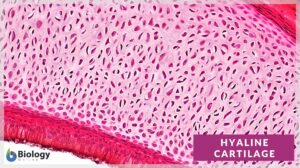
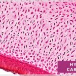
Definition: a type of cartilage that is characteristically glossy and smooth in appearance, and with interstitial substance containing fine type II collagen fibres obscured by the ground substance. Image shows the cross-section of hyaline cartilage (magnification: 200x).
Table of Contents
Hyaline Cartilage Definition
Before we define hyaline cartilage, let us understand what cartilage is. What is cartilage? Is it a connective tissue? Cartilage is a tough and flexible connective tissue in areas of high wear, such as bone ends, intervertebral discs, and joints. Is cartilage a bone? In certain animals including humans, the cartilage serves as the early skeletal framework at the embryonic stage. As the animal develops, most of it is replaced by bone. In cartilaginous fish (Chondrichthyes), the adult fish retains its cartilaginous skeleton. The cartilage tissues, compared to bone tissues, are more flexible and elastic.
Hyaline cartilage tissue (also referred to as hyaline connective tissue or hyaline tissue) is a type of a cartilage tissue. It is the most common type of cartilage characterized by a glossy and smooth appearance.
Where is hyaline cartilage found? Hyaline cartilage is found around the bones of free-moving joints. This is known as articular cartilage. Another example of hyaline cartilage is the tissue found in the walls of the respiratory tract. This includes the bronchi, the nose, the rings of the trachea, and the tips of the ribs.
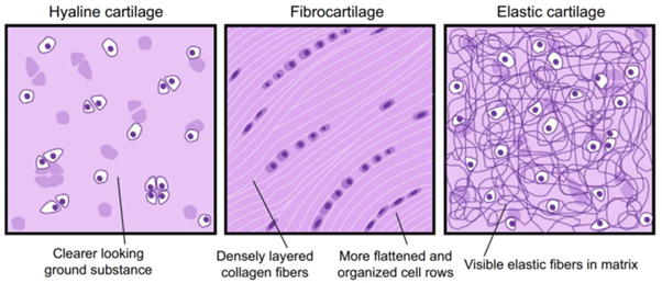
There are 3 different types of cartilage. So, apart from the hyaline cartilage, the other two are elastic cartilage and fibrocartilage. They differ mainly from the fibers that are present and will be discussed in more detail below.
The hyaline cartilage is a type of cartilage that characteristically has a shiny, white, semi-transparent appearance with a slightly blueish tinge. The word hyaline is derived from the Greek word ‘hyalos’, which means ‘glassy’ implying its shiny, smooth appearance. It is found in the larynx, trachea, and bronchi. Compare: elastic cartilage, fibrocartilage.
Hyaline Cartilage Structure
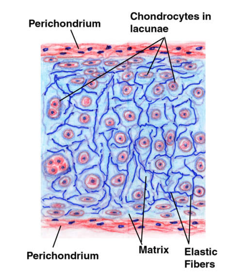
What is cartilage made of? Cartilage is made up of chondroblasts (or perichondrial cells) that produce the extracellular matrix (or ground substance), chondrocytes which lie in spaces known as lacunae, and collagen fibers. More details about the components of cartilage will be discussed below. See figure 2 for a hyaline cartilage diagram.
What is hyaline cartilage and where is it located?
How do you identify hyaline cartilage? It develops from mesenchymal cells, which are the stem cells found in the bone marrow. Hyaline cartilage does not contain any blood vessels or nerves so is a very simple structure. It obtains its nutrients via diffusion from nearby tissues.
Hyaline cartilage has a shiny, white semi-transparent appearance with a slightly blueish tinge. The word hyaline is derived from the Greek word ‘hyalos’ which means ‘glassy’ implying its shiny, smooth appearance. Interestingly, this appearance is lost as the tissue ages. In an embryo, hyaline cartilage makes up the first skeleton, then modifies as the embryo develops. This is by a process known as endochondral ossification.
Hyaline cartilage consists of fine type II collagen fibers, chondrocytes (matrix producing cells), and the extracellular matrix (or ground substance). Type II collagen fibers are thinner than type I collagen fibers. Type I, IV, V, VI, IX, and XI collagen are also present in very small quantities and help to strengthen the fibers together.
The cartilage matrix, known as the extracellular matrix or ground substance, is a gelatinous material abundant in glycosaminoglycans (GAGs), proteoglycans, and glycoproteins. The extracellular matrix fills the spaces between the cells and the fibers.
GAGs are essentially long polysaccharides made of amino sugars that attract sodium and potassium ions. These ions bring water along with it. Therefore, this helps to regulate the amount of water in the extracellular matrix.
Chondroitin sulfate and keratan sulfate are examples of sulfated GAGs and hyaluronic acid is an example of no non-sulfated GAG. These are all found in the extracellular fluid of cartilage.
Proteoglycans and glycoproteins are amino acids and carbohydrate molecules joined. They bind extracellular molecules and components together giving a gel-like fluid helping to absorb compression and force.
What are chondrocytes? Chondrocytes are the only cartilage cells to be found in hyaline cartilage. These cells start as chondroblasts (or perichondrial cells) which produce the cartilaginous matrix, then get immobilized within it in small spaces called lacunae.
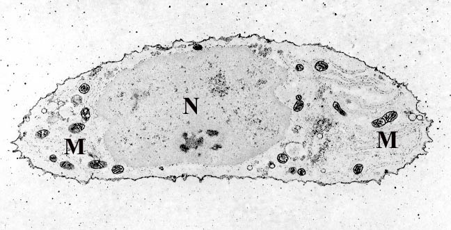
The role of chondrocytes is to develop, repair, and maintain the extracellular matrix. Chondrocytes have a limited healing capacity due to their limited replication ability. They rarely form cell-cell contact and are simply responsible for maintaining their immediate surroundings. Figure 3 shows the basic structure of a chondrocyte.
The hyaline cartilage is generally covered by the perichondrium. The perichondrium is found in developing bones but does not cover the articular cartilage on the ends of bones in adults. The perichondrium consists of an outer layer and an inner layer. The outer layer is fibrous cartilage and produces collagen fibers and the inner layer is involved in the formation of cartilage by forming chondroblasts or chondrocytes.
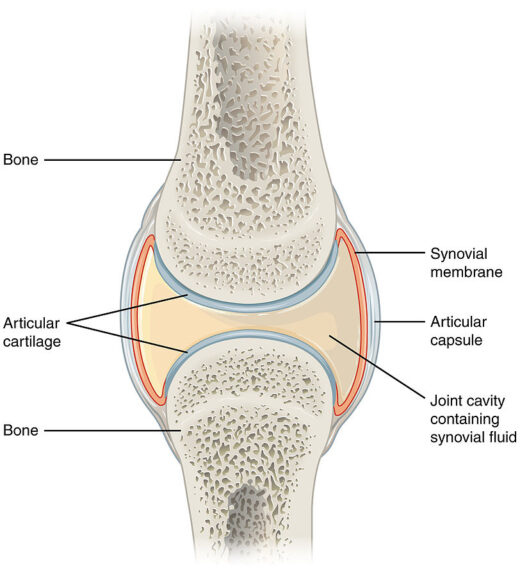
Hyaline Cartilage Example – Articular Cartilage
Articular cartilage is a type of hyaline cartilage. It is different from usual hyaline cartilage as it has flattened chondrocytes near the surface. In humans, it is 2 to 4 mm thick. It does not have blood vessels, nerves, or lymphatics. Its ECM is dense whereas chondrocytes are sparse.
Deeper in the tissue, the chondrocytes take a more typical structure. In the very deep layers of the cartilage, the cells are found in columns with a calcified matrix. The collagen fibers form arches giving it a strong structural arrangement to withstand pressure.
Articular cartilage is composed of type II collagen but has also been found to contain small amounts of type VI, IX, X, and XI collagen. Figure 4 shows the location of the articular hyaline cartilage in a joint.
Articular cartilage is made up of different zones. These include the superficial zone, followed by the middle transitional zone, the deep zone, and finally the calcified zone. Within each zone, there are 3 regions. These are the pericellular region, the territorial region, and the interterritorial region. The video below presents the layers found in articular cartilage.
The superficial zone makes up around 10-20% of the total thickness of the cartilage. Collagen fibers II and IX can be found here. It contains a large volume of chondrocytes that have a more flattened appearance.
The superficial zone is in direct contact with the synovial fluid and protects the deeper layers from force and stresses.
The middle zone follows directly from the superficial zone and provides the bridge to the deeper layers. This zone represents around 40-60% of the total cartilage thickness. It consists of thicker collagen fibers and proteoglycans. The chondrocytes here are spherical and found in small amounts.
The middles zone functions to protect against compacting forces. The deep zone follows on from the middles zone and provides the best resistance to compacting forces. It contains the highest proteoglycan content and the least water content. The collagen fibers are arranged at right angles to the surface and the chondrocytes are arranged in columns. It represents around 30% of the total articular cartilage volume. Finally, the calcified zone attaches the cartilage to the bone. It does this by anchoring the collagen fibers in the deep zone to the subchondral bone.
Hyaline Cartilage Histology
As mentioned above, hyaline cartilage connective tissue is made up of cells and fibers within an extracellular matrix. Hyaline cartilage histology describes how hyaline cartilage looks when viewed under a microscope.
The chondrocytes can be seen as rounded or angular in form. In adult cartilage, the cells are present in isogenous groups, formed from a single progenitor cell. The matrix is visually homogenous and basophilic in appearance. The reason for this is because of the high concentration of sulfated GAGs in the matrix mask the collagen fibers. Type II collagen fibers are also very small which is why the extracellular matrix appears so shiny and smooth.
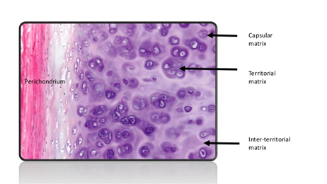
There is no uniform distribution within the extracellular matrix. Therefore, the three basic zones can be seen. Figure 5. Shows these different zones.
- The capsular matrix, which consists of a thin zone that surrounds each lacuna. Here lies the highest concentration of sulfated GAGs.
- The territorial matrix, which surrounds the capsular matrix.
- The interterritorial matrix, which is less basophilic due to a lower concentration of sulfated GAGs and a higher proportion of collagen.
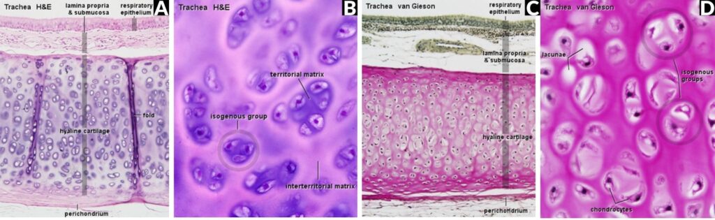
The hematoxylin and eosin (H&E stain) along with the Van Geison staining methods can both be used to look at hyaline cartilage under the microscope. The Van Geison stain uses picric acid and acid fuchsin and stains collagen red. The cartilage is viewed as a red zone lying below the epithelium. The staining is lighter where it becomes closer to the lacunae indicating the territorial matrix.
In H&E stained sections, the color intensities are reversed, but give a better definition than with the Van Geison stain. The territorial matrix is dark, and the interterritorial matrix is a lot lighter in color. Groups of chondrocytes can be found surrounded by these darker areas in the H&E method. These chondrocytes are derived from the same progenitor and are therefore an isogenous group. The perichondrium surrounds the cartilage except in the articular cartilage.
Figure 6 images show Van Gieson, then H&E staining of the trachea.
The Difference from Other Types of Cartilage
There are 3 different cartilage types found in the body. Hyaline cartilage is the most common but also the weakest type of cartilage. The other two types of cartilage are fibrocartilage and elastic cartilage. How is hyaline cartilage different from elastic cartilage or fibrocartilage? See below to see descriptions of each cartilage type. The visual differences between the types of cartilage can be seen in Figure 1.
Elastic Cartilage
Let us look at Hyaline cartilage vs elastic cartilage. Elastic cartilage (or yellow fibrocartilage) provides strength and elasticity to certain parts of the body. Where is elastic cartilage found? It can be found in the external part of the ears known as the pinna and the epiglottis and laryngeal cartilages and auditory tube/eustachian tube. Elastic cartilage functions to give support with additional elasticity. It contains a dense network of elastin fibers. It does not protect from mechanical stress or compression. More details such as elastic cartilage locations and appearance are highlighted in table 1 below.
Fibrocartilage
Fibrocartilage connective tissue is a dense flexible and supporting cartilage made up of fibrous tissue. Where is fibrocartilage found? Fibrocartilage locations include the intervertebral discs of the spine, in the jaw, and the knee and wrist. This fibrocartilage tissue contains large bundles of type I collagen. It is the strongest type of cartilage. Fibrocartilage functions to give support against weight-bearing and pressure forces.
Fibrocartilage can be described by separating it into 4 different groups.
- The first group is intra-articular fibrocartilage. This acts as a buffer in between joints that are exposed to high impact and frequent movement. An example is the menisci of the knee.
- The second group is connecting fibrocartilage which is found in joints with limited motion such as the intervertebral discs.
- Stratiform fibrocartilage is found coating the bone grooves where tendons and muscles lie.
- Finally, the last group, circumferential fibrocartilage surrounds some articular cavity margins which protect their edges. An example is an acetabular labrum (lining the hip socket).
Fibrocartilage vs hyaline cartilage, as well as the locations in the body where fibrocartilage is located, is described in table 1 below.
| Table 1. Hyaline cartilage vs Elastic Cartilage and Fibrocartilage | |||
|---|---|---|---|
| Cartilage – Types | Hyaline Cartilage | Elastic Cartilage | Fibrocartilage |
| Appearance | Translucent and glossy | Glossy and yellow | White, dense, and opaque |
| Location | Fetal skeleton until maturation
The growth plate at the end of long bones Costal Cartilage at the end of the ribs Nose Larynx Bronchi Articular cartridges |
The outer ear (pinna)
Epiglottis External Auditory tube/eustachian tube Laryngeal cartilages |
Intervertebral discs
Pubic symphysis joint Articular discs in sternoclavicular and temporomandibular joints The glenoid labrum in the shoulder blade The acetabular labrum in the hip joint |
| Main Collagen Type | Type II | Type II | Type I |
| Chondrocytes | Small, arranged in groups of 2 – 8 cells | Large, arranged in groups of 2 – 4 cells | Small, between bundles of collagen fibers. Arranged in strips |
| Extracellular Matrix | Homogenous and basophilic. | High in elastic fibers. | High in collagen fibers. Eosinophilic. |
| Perichondrium | Present | Present | Absent |
Hyaline Cartilage Function
So, what is the function of hyaline cartilage? Hyaline cartilage contains relatively few fibers and provides a smooth surface for movement as well as a cushion that absorbs shock where the bones meet. In articular cartilage, the primary function is to provide a smooth surface that can withstand friction and pressure from weight-bearing functions.
In the trachea, it provides support for the softer tissues and allows them to maintain an open position.
Biological Importance of Hyaline Cartilage
The most important role of hyaline cartilage is to provide mechanical support for the respiratory system, developing bones and articular surfaces.
As we age, problems with the quality of our hyaline cartilage can arise. With increased age, the number of chondrocytes in the superficial layer of articular cartilage drops whereas the number of chondrocytes in the deeper layers increases. Also, with an increase in age is a decrease in proteoglycans in the extracellular matrix. There is also an increase in keratin sulfate and a decrease in chondroitin sulfate. The volume of hyaluronic acid also increases. Hyaline cartilage is susceptible to wear and tear due to its role as a shock absorber and heavy use in daily activities. All these factors can lead to hyaline cartilage becoming more susceptible to damage and disease than the other types of cartilage.
Cartilage tissues are likely to be slow in healing following an injury because there is a lack of blood supply to the chondrocytes. This means that the matrix is slow to form. Also, chondrocytes are stuck in lacunae and cannot migrate to an area of damaged tissue. Damaged tissue becomes scar tissue.
In the extracellular matrix, chondroitin sulfate plays an important part as it is an anti-inflammatory mediator and reduces pain. Some studies suggest its presence helps slow the break-down of cartilage and thereby prevents conditions such as osteoarthritis. Osteoarthritis occurs when the cartilage wears away, allowing the bones to rub against each other causing subchondral bone (bone just below the cartilage) sclerosis (hardening), and inflammation of the synovial membrane leading to pain.
Hyaline Cartilage in Other Animals
Animals in the class Chondrichthyes have a skeleton composed completely of cartilage. Sharks and rays are good examples of this. Cartilage is less dense than bone, yet still provides strength and therefore allows these animals to move quickly through the water without exerting too much effort.
Cartilage is also found in invertebrates, such as horseshoe crabs, snails, and cephalopods (predatory mollusks e.g. octopus and squid). The branchial cartilage in the arthropod Atlantic horseshoe crab (Limulus polyphemus), is rich in vacuolated chondrocytes that differ from any other arthropod.
Endosternite cartilage is another type found in this species. It is more fibrous than the hyaline cartilage found in vertebrates. It is found near the ventral nerve cords and gill cartilage tissue.
In the octopus (an example of a cephalopod), the cranial cartilage resembles hyaline cartilage and is one of the only hard parts of the octopus body. The growth of this cartilage occurs via cells moving from the outside to the center. In the common cuttlefish (Sepia officianalis), the cartilage is fibrillar collagen. The growth pattern of this cartilage is essentially the same as in vertebrate cartilage.
In gastropods (snails, slugs, or whelks), the odontophore is a cartilage-formed feeding structure that provides feeding support. The odontophore is cell-rich cartilage containing myoglobin that is surrounded by a low volume of extracellular matrix and collagen.
Finally, in the feather duster worms (Sabellid polychaetes), cartilage supports their tentacles.
Conclusion
Overall, cartilage can be described as a vital structural part of the body found in vertebrates and some invertebrates. It is a firm but soft tissue that provides support, flexibility, and strength. The importance of cartilage can be seen in hyaline cartilage between joints. Here, as we age, the cartilage thins leading to inflammation and bone rubbing. Studies are ongoing by researchers in this area of science that can help to improve our understanding of the processes that lead to this and develop ways to counteract/treat/prevent such diseases.
Try to answer the quiz below to check what you have learned so far about hyaline cartilage.

References
- Bengochea, K. (2020). Hyaline Cartilage histology. KenHub.
- cartilage | Description, Anatomy, & Function | Britannica. (2020). In Encyclopædia Britannica. https://www.britannica.com/science/cartilage#ref213485
- Cartilage. Anatomy made Simple. https://anatomyqa.com/cartilage-histology/
- Cartilage. SBPMD Histology Laboratory manual. http://www.columbia.edu/itc/hs/medical/sbpm_histology_old/lab/lab06_cartilage.html
- Cartilage: The Three Types of Cartilage. Histology Guide © Faculty of Biological Sciences, University of Leeds. https://www.histology.leeds.ac.uk/bone/cartilage_types.php
- Henrotin, Y., Mathy, M., Sanchez, C., & Lambert, C. (2010). Chondroitin sulfate in the treatment of osteoarthritis: from in vitro studies to clinical recommendations. Therapeutic advances in musculoskeletal disease, 2(6), 335–348. https://doi.org/10.1177/1759720X10383076
- Horner, C.B., Low, K., Nam, J. (2016). 10 – Electrospun scaffolds for cartilage regeneration. Nanocomposites for Musculoskeletal Tissue Regeneration. 213-240. https://doi.org/10.1016/B978-1-78242-452-9.00010-8
- Kidd, G. Difference between hyaline cartilage and elastic cartilage. Difference Between.net. http://www.differencebetween.net/science/health/difference-between-hyaline-cartilage-and-elastic-cartilage/
- Maynard, R.L. Downes, N. (2019). Chapter 3 – Introduction to the Skeleton: Bone, Cartilage and Joints. Anatomy and Histology of the Laboratory Rat in Toxicology and Biomedical Research. (3) 11-22. https://doi.org/10.1016/B978-0-12-811837-5.00003-4
- Slomianka, L. Blue Histology Skeletal Tissues – Cartilage. School of Anatomy and Human Biology – The University of Western Australia. https://www.lab.anhb.uwa.edu.au/mb140/CorePages/Cartilage/Cartil.htm#:~:text=Types%20of%20Cartilage-,Hyaline%20Cartilage,connective%20tissue%2C%20from%20mesenchymal%20cells.&text=Interstitial%20growth%20%2D%20Chondroblasts%20within%20the,a%20thin%20partition%20of%20matrix.
- Sophia Fox, A. J., Bedi, A., & Rodeo, S. A. (2009). The basic science of articular cartilage: structure, composition, and function. Sports health, 1(6), 461–468. https://doi.org/10.1177/1941738109350438
- The truth about Glucosamine and Chondroitin Sulphate. Orthopedic Associates. https://www.oaph.com/patient-resources/education/truth-about-glucosamine-and-chondroitin-sulfate
- Watkins, J., Mathieson, I. (2009). Connective Tissues. The Pocket Podiatry Guide: Functional Anatomy. (4) 107-156. https://doi.org/10.1016/B978-0-7020-3032-1.00004-4
©BiologyOnline. Content provided and moderated by BiologyOnline Editors.