Cell membrane
n., plural: cell membranes
[sɛl ˈmembɹeɪn]
Definition: The cell’s outer membrane is made up of two layers of phospholipids with embedded proteins and separates the contents of the cell from its outside environment, as well as regulates what enters and exits the cell
Table of Contents
Cell Membrane Definition
Just like any non-living body possesses a plastic or paper packaging material that keeps the contents of the body intact, in shape, protected, and well-preserved, the cells have a protective outer layer called the “Cell membranes (CM)” or “Plasma membrane (PM)” or cytoplasmic membrane. Whether it be a prokaryotic cell or eukaryotic cell, the presence of a cell membrane has been noticed in all.
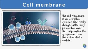
What is a cell membrane? Basically, a cell membrane (or plasma membrane) is an ultrathin, plastic, dynamic, electrically charged, and selectively-permeable membrane layer that separates the cytoplasm from the extracellular matrix and helps in maintaining the cell structure and function. This should not be confused with the cell wall, which is an extra layer present outside of the cell membrane mainly in plants, bacteria, and fungi.
An animal cell membrane is an outermost layer whereas the plant cell membrane is the second layer after the plant cell wall. Being selectively permeable, the cell membrane allows the movement of both solvent and some selected solutes. The movement is along the concentration gradient.
We should take note here that the membrane is selectively permeable and not semi-permeable. Semi-permeability means that a membrane will allow the movement of solvent only from its higher concentration to lower concentration; no solute movement is permitted. Look at the cell membrane diagram below to have a basic idea of its position inside a cell.
The cell membrane is a membrane that surrounds the cell and separates it from the outside environment. In animals, this membrane is the outermost covering of the cell whereas in plants, fungi, and some bacteria it is located beneath the cell wall. Although some cells form another layer above the cell membrane (called cell wall), other cells have the cell membrane as the only protective barrier between the cytoplasm and the outside of the cell. Synonyms: plasma membrane; cellular membrane; cytoplasmic membrane; plasmalemma.
Now that we know how to define cell membranes, let’s move ahead and understand how the idea of membrane structure evolved over the years.
There are different cell membrane models that help us develop further clarity about its structure, function, roles, and purposes in a biological system.
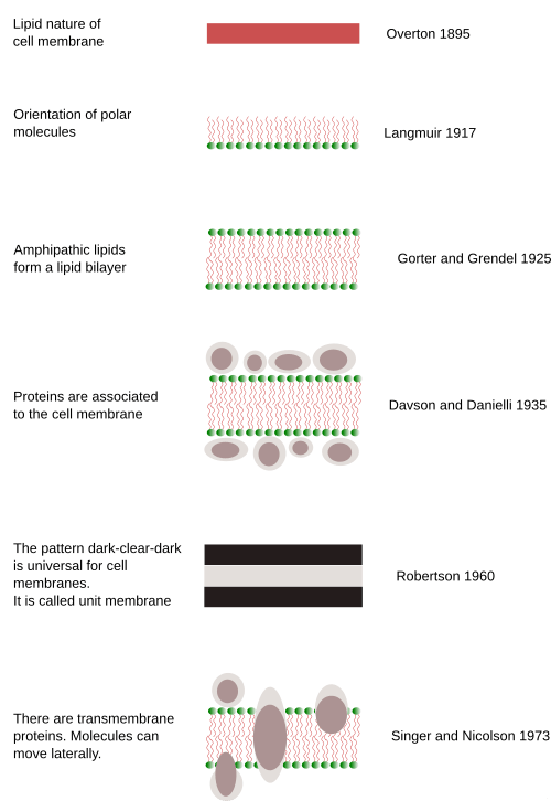
1. Overton Model
- Came in 1910
- The first model to explain the structure of plasma membrane
- Early on when there was no clear idea about what makes up cell membranes; Overton implied that PM is coated with lipids-soluble material.
- He also suggested that non-polar solutes can easily cross the membrane but polar solutes can’t cross the membrane at all; and are therefore discarded!
2. Irving Langmuir Model
- Came in 1925
- The first model to say that the cell membrane is made up of monolayers
- They are made up of a single layer of amphipathic phospholipid molecules in which the hydrophobic tails protrude away from water and hydrophilic heads face the water.
3. Gorter and Grendel Model
- Came in 1924
- The first model to say that components of the cell membrane are various types of lipids, but are double layered.
4. Davson and Danielli Model
- Came in 1935
- This is a trilaminar model or the sandwich model that tried to explain the presence of proteins in the plasma membrane for the first time apart from the major phospholipid functions in the membrane.
- It implied that the membrane is a protein-lipid-protein sandwich.
5. Robertson Model (Unit Membrane Model)
- Came in 1950s
- It explained that there’s no space between the phospholipid bilayers.
- It also implied that the thickness of the membrane is measurable.
- By performing experiments with different types of cells, he came to another conclusion that the basic underlying structure of the cell membrane is the same- hence called his model the “Unit membrane model”.
6. Singer and Nicholson Model (Fluid Mosaic Model)
- The most accepted model of the membrane structure till today.
- Came in 1972
- Definition of Fluid mosaic model: The model explains that proteins are mosaics in the fluid-like lipid part of a membrane, indicating that the proteins aren’t present in a layer but in some places in a discontinuous manner.
- It explains that the plasma membrane is composed of both lipids and proteins but proteins in lipids are analogous to icebergs in the sea.

Now, we can hope that a better picture of the cell membrane must have developed in your mind. The different models help us understand how Science and scientists interpret the biological systems in varied ways and how the basic concept evolves with rational model building over years…
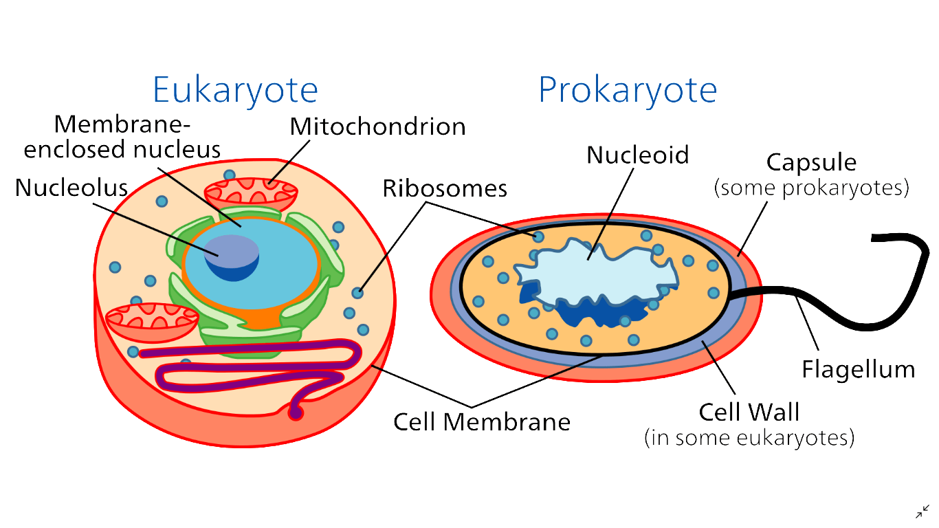
To understand where is the cell membrane located in both prokaryotic and eukaryotic cells, look at the figure below.
Now let’s move on to the structure of the cell membrane and get to know what are the different constituents of a membrane.
Cell Membrane Structure
When talking about the structure of a cell membrane, the important thing to learn is the answer to this question, what is the cell membrane made of? The cell membrane is composed of varying amounts of lipids, carbohydrates, and proteins. Although lipids form the basic structure of the plasma membrane, proteins and carbohydrates play irreplaceable roles in the functioning of biological membranes.
Lipids
Membrane lipids are amphipathic in nature. So what are lipids made of? They are made up of hydrophilic polar heads and hydrophobic non-polar fatty acid tails. There are mainly 3 types of lipids present in the plasma membrane.
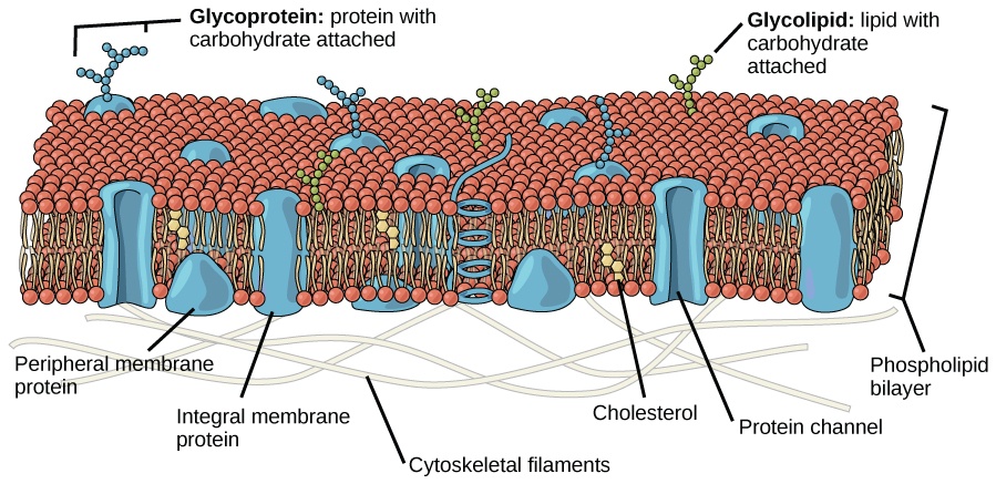
1) Phospholipids – Phospholipid molecules are amphipathic lipids with a phosphate group attached with a covalent bond. They are the most abundant form of lipids present in the cell membrane often constituting more than 50% of the total lipids. They are arranged in two layers with the hydrophilic ends in contact with the cytosol of the cell & with the extracellular environment. The hydrophobic ends of both layers form the core of the cell membrane. Some lipids examples are glycerophospholipids (major component) and sphingophospholipids (minor). So, when asked what is a phospholipid, we can tell it’s a major type of lipid found in cell membranes.
2) Glycolipids – They are lipids with carbohydrates attached with a glycosidic bond. They are present in minute amounts and constitute only about 2% of the total lipids of the cell membrane. However, they play a crucial role in maintaining the stability of the cell membrane & in cellular recognition. It is interesting to note that the different blood groups in humans are determined by the oligosaccharide group of the glycolipids on RBC’s membrane. Some common glycolipids are globoside, cerebroside, ganglioside, etc.
3) Sterols – The rest of the remaining lipids are sterols. Cell membranes of the plants usually contain sterols and that of animals contain cholesterol. They both serve the similar purpose of regulating the fluidity of the membrane at different temperatures.
For example, cold-blooded animals contain the maximum amount of cholesterol in cell membranes that act as an antifreeze agent. However, at high temperatures, the cholesterol reduces the movement of fatty acid chains, and therefore, reduces the fluidity and reduced permeability of the cell membrane. In plants, the same role is played by the sterols.
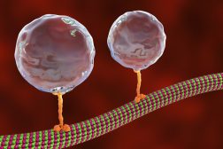
Phospholipids forming lipid vesicles
Many times, lipids form vesicles called liposomes. Liposomes are different from micelles as liposome formation occurs from chiefly glycerophospholipids. On the other hand, micelle formation happens from sphingophospholipids. Liposomes are spherical in structure and happen to have a planar bilayer structure while micelles are single-layered.
Carbohydrates
Membrane carbohydrates are mostly present in the form of glycolipids, glycoproteins & proteoglycans. The carbohydrate part is present mostly outside of the cell surface. This makes up a loose carbohydrate coat present outside of the cell membrane known as glycocalyx.
The carbohydrates serve the following important functions:
- Repels the negatively charged particles as some carbohydrate moieties are themselves negatively charged.
- Attachment to the adjacent cells by the interaction between the glycocalyx.
- Cell surface receptors. For e.g. Insulin receptors.
- Play a role in immune reactions.

Proteins
Proteins make an important contribution to the cell membrane, which constitutes around 50% of the total membrane volume. There are mainly 3 types of membrane proteins.
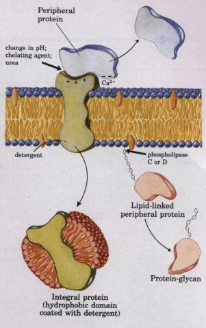
- Integral membrane proteins – These proteins span the entire width of the cell membrane. They have 2 hydrophilic domains that interact with the intracellular and extracellular environment respectively and 1 hydrophobic domain that anchors the protein within the core of the cell membrane.
Examples – Ion channels, proton pumps, G-protein coupled receptors. - Lipid anchored proteins – These proteins are characterized by their covalent attachment to the membrane lipid. These proteins can be found on either side of the membrane.
Example – G protein - Peripheral membrane proteins – These proteins interact with the membranes transiently. They either attach to the integral membrane proteins or the periphery of the lipid bilayer. Once reacted, the protein molecule dissociates to carry on its own function within the cytosol.
Example – Hormones, Enzymes
Cell Membrane Functions
Moving on to the vital roles and functions the cell membrane serves in the biological functioning of a cell, we can decipher the following points. What is the function of the cell membrane in the biological world?
1) Shape & Structure of the cell – The cell membrane acts as an anchor to the cytoskeleton.
2) Barrier and gatekeeper of the cell – The cell membrane protects the cytosol from the external environment.
3) Cellular transport – It regulates the molecular transport of substances across the membrane.
4) Intercellular junctions – Gap junctions, tight junctions, etc.
5) Cell communication and recognition – help tissues work together in unison.
6) Cell signaling – contains receptors and enzymes.
So, when asked what does the cell membrane does and why is the cell membrane important, we can reiterate these points.
Common biological reactions
Cell recognition is one of the ways by which cells communicate with one another. It is possible through specific cell adhesion molecules on the surface of the cell. An example of cell recognition is integrin (LFA-1) of T cell binding to ICAM of endothelial cell. Another is selectin (L) of lymphocyte binding to addressin (CD34) of endothelial cell.
One of the major functions of the cell membrane is transport. The cell membrane is involved in both the passive and active types of transport. In passive transport, substances move along the concentration gradient. This is in contrast to active transport, which is a type of transport characterized by an uphill movement of substances (i.e. from lower to higher) and therefore requires chemical energy, e.g. ATP. In moving substances across a biological membrane, a passive transport may or may not need the assistance of a membrane protein.
READ: Movement of Molecules Across Cell – Tutorial
There are four major types of passive transport are (1) simple diffusion, (2) facilitated diffusion, (3) filtration, and (4) osmosis. Simple and facilitated diffusions refer to the net movement of molecules from higher to lower concentrations. Osmosis refers to the diffusion of a solvent (usually water molecules) through a semipermeable membrane from lower to higher solute concentrations. Filtration is the movement of water and solute molecules across the cell membrane driven by hydrostatic pressure that is generated by the cardiovascular system.
Endocytosis is the process in which a cell takes in materials (e.g. proteins and hormones) from the outside by engulfing and fusing them with its plasma membrane. There are two main types of endocytosis: phagocytosis, which literally means cell-eating, and pinocytosis, which literally means cell-drinking.
The cell engulfs by creating a small deformation inward (invagination) containing the substance to be transported inside the cell. The invagination is then pinched off from the cell membrane, resulting in a vesicle containing the substance. Since endocytosis requires ATP, it is considered a form of active transport.
Exocytosis is the process in which the cell seems to spit out materials from the cell. Thus, exocytosis seems the opposite process of endocytosis. The vesicle containing the material fuses with the cell membrane and then the contents are extruded outside the cell into the surrounding medium.
Biological importance
The structure and composition of the cell membrane make it selectively permeable (or semipermeable), which means not every substance is allowed to enter or leave the cell. The cell membrane controls which substances can go in and out of the cell. It can allow a particular substance to pass through at a certain time and then reject the same substance at a later time. The presence of surface molecules (e.g. glycoproteins, glycolipids, etc.) serves as the ‘signature’ of a cell. Every cell has a different ‘signature’ or ‘marker’ that is thought to function in cell recognition, or in a sort of cellular identification system. Its other main functions include cell adhesion, ion channel conductance, cell signaling, and attachment point for cytoskeleton (which is important in keeping the shape of the cell).
Prokaryotes
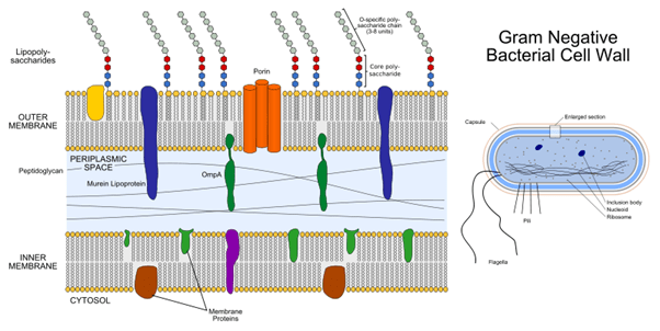
Both prokaryotes and eukaryotes possess cell membranes. Prokaryotes are majorly studied under 2 categories, namely Archaea and Bacteria. Unlike eukaryotes which have a cell membrane and membrane-bound organelles, prokaryotes only have one cell membrane but no membrane around their organelles. Additionally, among bacterial prokaryotes, two types emerge; they are gram-positive bacteria and gram-negative bacteria. Gram-negative bacteria have an additional outer membrane apart from the cell membrane.
Structure
Now let’s put a little light on some interesting structural intricacies of the cell membrane.
Fluid mosaic model
The fluid mosaic model was given by SJ Singer and G. L. Nicolson. This is the most acceptable model of the cell membrane. This model describes the cell membrane as a 2-dimensional fluid that restricts the lateral diffusion of the membrane components. The diffusion across a biological membrane is called simple diffusion. According to it, the main function of the cell membrane is to separate the internal contents of the cell from the outside.
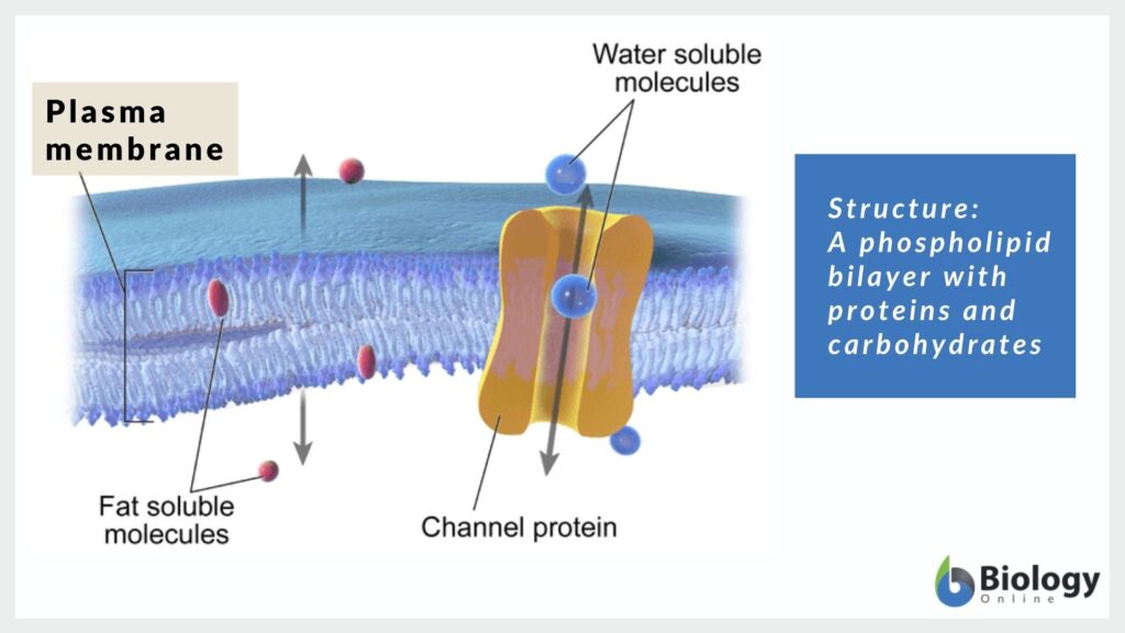
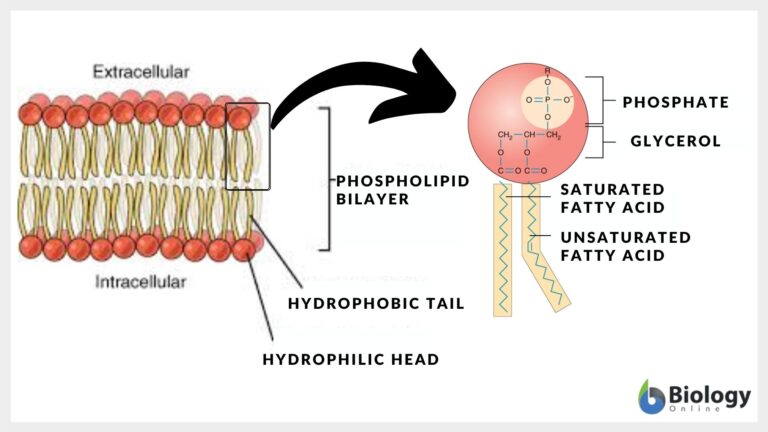
Lipid bilayer
The cell membrane is arranged in two layers of amphipathic membrane phospholipids. Each layer is one molecule thick and spans over the entire cell. One end of the phospholipid contains the phosphate group which is the hydrophilic part and the other end of the phospholipid contains fatty acids which is the hydrophobic part. (Meaning of hydrophobic is anything that repels water; hydrophobic substances like salad oil are also water-repelling in nature).
The hydrophilic parts of each layer repel each other, and the hydrophobic parts of each layer tend to interact with each other. Hence, the hydrophilic parts interact with the inside and the outside of the cell. The core of the cell membrane is made up of the hydrophobic ends of both the lipid bilayer.
This arrangement keeps the water-soluble substances from moving across the cell membrane. However, lipid-soluble substances such as carbon dioxide, alcohol can easily cross the cell membrane.
Membrane polarity
Membrane polarity is the asymmetric distribution of the proteins within different domains of the cell membrane. For example, in epithelial cells, the basal and lateral surfaces are identical in composition and activity, but it is distinctly different from the apical surface. This asymmetry may be explained by the presence of tight junctions near the apical surface that would not allow the migration of ion channels and other embedded proteins from the basolateral to the apical surface.
Membrane structures
There are numerous membrane structures that help in the basic cellular processes. They are as follows:
1. Adhesion
2. Molecular movements, e.g., endocytosis and exocytosis
(If asked, “does endocytosis require energy?” The answer is YES, in the form of ATP)
(Also, if asked, “does exocytosis requires energy?” The answer is again YES!)
3. Cell-to-cell communication
4. Cell junction activities
5. Signal transduction (via Caveolae and Postsynaptic density)
6. Cellular motility and invasion (via Podosomes)
7. Interactions between extracellular matrix and cell (via Focal Adhesion)
8. Permeability barriers across membranes (Cell junctions)
Cytoskeleton
The cytoskeleton of the cell extends from the cell membrane to the nucleus. It is made up of microfilaments, intermediate filaments, and microtubules which can grow or disassemble depending on the requirement of the cell.
Intracellular membranes
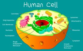
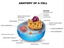
Now as we have learned that prokaryotes don’t have membranes around their organelles (not even around the nucleus- hence called nucleoid), eukaryotic cells have membranes around their organelles. Some organellar parts of the cell are single membrane-bound whereas others are double membrane-bound. Mitochondria, chloroplast , and nucleus possess two membranes: one outer and one inner. On the other hand, endoplasmic reticulum, Golgi bodies, peroxisomes, lysosomes, and vacuoles have just one membrane.
Variations
The ratios and percentages of the different components vary from cell to cell. For example:
· RBC Membrane: 52% Proteins, 40% Lipids, 8% Carbohydrates
· Neuron Membrane: 18% Proteins, 73% Lipids, 9% Carbohydrates
· Thylakoid Membrane: 70% Proteins, 30% Lipids, 0% Carbohydrates
· Normal Plant Cell Membrane: 40% Proteins, 40% Lipids, 20% Carbohydrates
Permeability
The cell membrane is selectively permeable and only allows movement of certain specific substances across the cell membrane which are necessary for its survival. The cell does this by various transport mechanisms that can be either passive (without the use of ATP/energy) or active (with the use of ATP/energy)
Conclusion
By learning the cell membrane’s structure, functions, variations in composition, and specialization, we can, at last, conclude how pivotal a role is played by it. Without a cell membrane, the integrity and the biological functioning of a cell are unimaginable.
Interesting Fact
1. Cholesterol (in the plasma membrane of animal cells)
2. Ergosterol (in the plasma membrane of fungal cells)
3. Sitosterol and Stigmasterol (in the plasma membrane of plant cells)

But prokaryotic cells interestingly lack any such form…. They possess “hopanoids” but hopanoids don’t have a steroid nucleus (have “five fused rings”)
Even in prokaryotes, an interesting thing that has been noticed is that the plasma membrane of Mycoplasma has a sterol type called cholesterol, which is usually found in the plasma membrane of animal cells.
Try to answer the quiz below to check what you have learned so far about cell membranes.

References
1. Casares, D., Escribá, P. V., & Rosselló, C. A. (2019). Membrane Lipid Composition: Effect on Membrane and Organelle Structure, Function and Compartmentalization and Therapeutic Avenues. International journal of molecular sciences, 20(9), 2167. https://doi.org/10.3390/ijms20092167
2. Cooper GM. (2000) The Cell: A Molecular Approach. 2nd edition. https://www.ncbi.nlm.nih.gov/books/NBK9898/
3. Singer, Jonathan S., Nicolson G.L. (1972) The fluid mosaic model of the structure of cell membranes.” Science 175.4023: 720-731.
4. Raicu, Valerica, and Aurel Popescu. (2008). Cell Membrane: Structure and Physical Properties.” Integrated Molecular and Cellular Biophysics 73-99.
5. Yeagle P.L. (2016) The Membranes of Cells Book, Third Edition
6. Buehler L. (2015) Cell Membranes, First Edition
7. Helgi I. Ingólfsson, Manuel N. Melo, Floris J. van Eerden, Clément Arnarez, Cesar A. Lopez, Tsjerk A. Wassenaar, Xavier Periole, Alex H. de Vries, D. Peter Tieleman, and Siewert J. Marrink. (2014) Journal of the American Chemical Society. 136 (41), 14554-14559. DOI: 10.1021/ja507832e
8. Harayama, T., Riezman, H. (2018) Understanding the diversity of membrane lipid composition. Nat Rev Mol Cell Biol 19, 281–296 (2018). https://doi.org/10.1038/nrm.2017.13
© Biology Online. Content provided and moderated by Biology Online Editors