
noun,
plural: pharynges or pharynxes
[ˈfæɹ.ɪŋks]
Definition: The cavity at the back of the mouth
Table of Contents
Lined with moist tissue, the pharynx is like a bustling biological highway that directs food and air to their respective destinations in the body. With unique functions allotted to its different regions, the pharynx anatomy is equally spectacular. While most of us are only aware of its common conduit roles for food and air, we’ll enlighten you about its role in human speech and immunity in this article. Read on to learn more about this anatomical marvel that plays a vital role in our daily lives while we often take its many functions for granted.
Pharynx Definition
The pharynx can be defined as a biological cavity lined with moist tissue that connects the mouth (oral cavity) and the nostrils (nasal cavities) to the esophagus (food pipe), trachea (windpipe), and larynx region.
With both respiratory as well as digestive functions, the pharynx forms an indispensable part of two major organ systems; the respiratory tract and digestive systems. The pharynx marks its presence in both categories of animals namely; vertebrates and invertebrates.
By its nature, the pharynx is:
- Shape: Cone-shaped hollow structure
- Layers: Surrounded by moist tissue on all sides
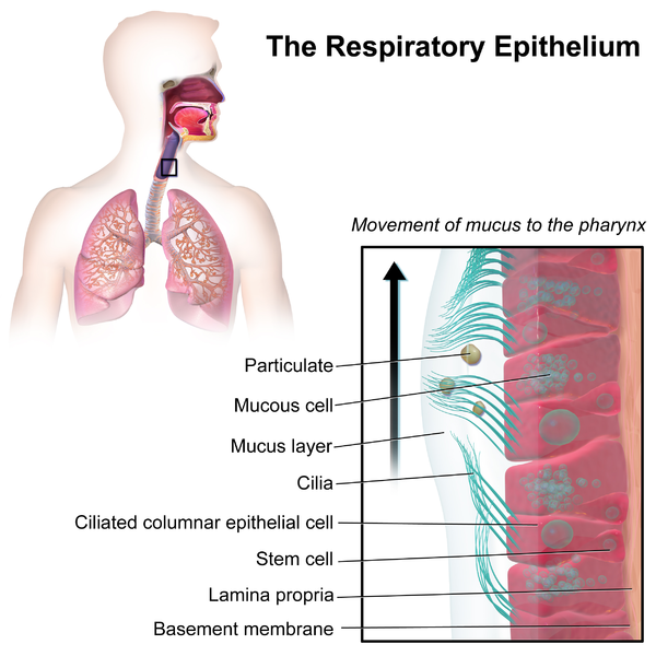
- Role: Serves as a mucus-rich barrier
- Muscular in nature: Muscles of the pharynx are thick and fibrous type with both circular and longitudinal variations.
- Circular muscles: Role in pushing food down the esophagus and in the prevention of swallowing air.
- Longitudinal muscles: Role in lifting the pharyngeal walls during swallowing.

Figure 1: Mucus layers in the respiratory epithelium. Image Credit: BruceBlaus.
Watch this vid about
Biology definition:
The pharynx is the ultimate multi-tasker of the body, acting as a crucial passageway for food and air. It’s a muscular tube that connects the mouth and nose to the esophagus, trachea, and larynx, allowing us to breathe, swallow, and speak.
Etymology: From Ancient Greek φᾰ́ρῠγξ (phárunx), meaning throat.
Pharynx Location
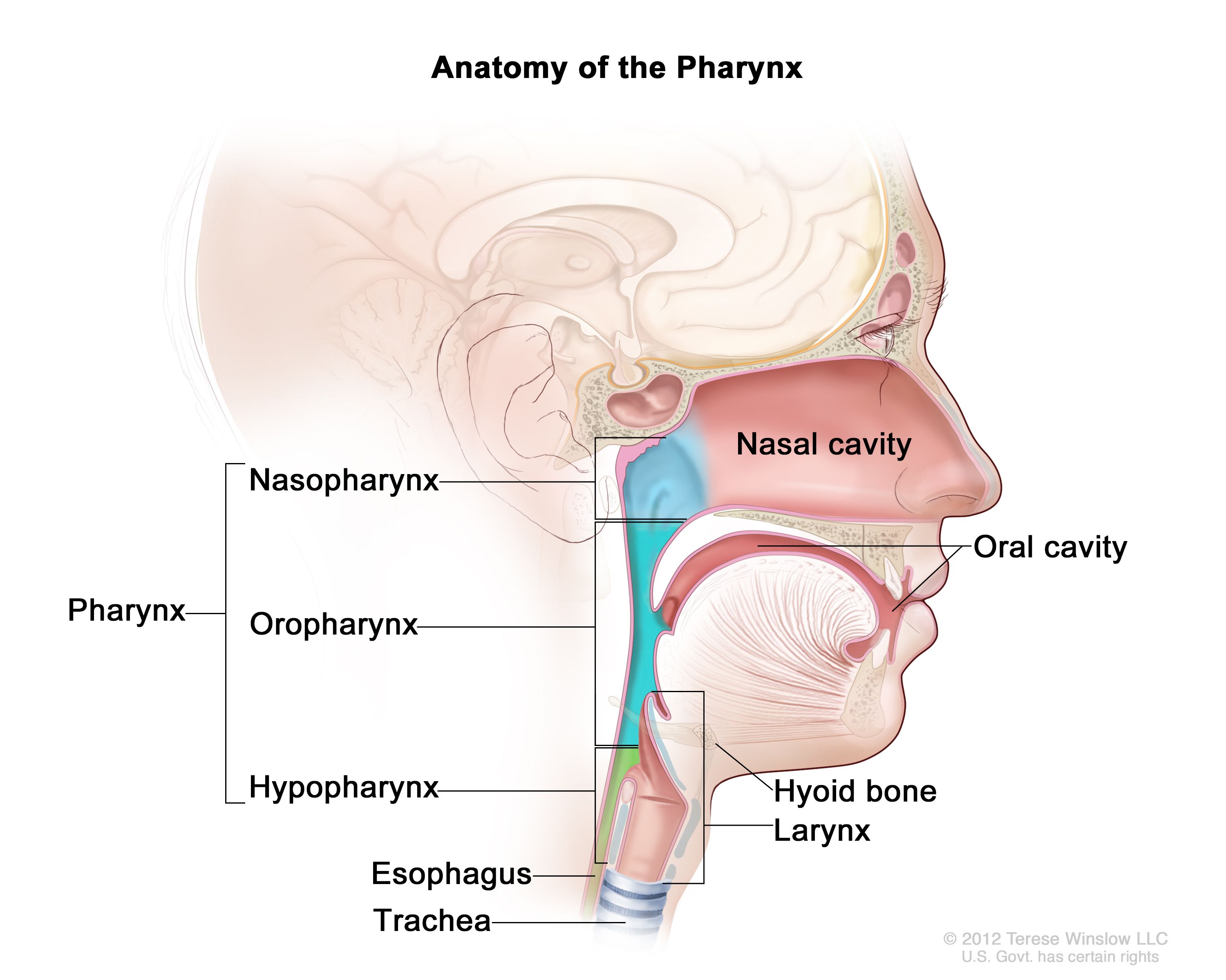
When we discuss the location of the pharynx, we must account for its 3 major divisions.
- Nasopharynx or nasal pharynx (part of pharynx connected to the nasal cavity)
- Oropharynx or oral pharynx (part of the pharynx connected to the oral cavity)
- Laryngopharynx or hypopharynx (part of the pharynx connected to the larynx and food pipe [esophagus])
So, when explaining the precise location of the pharynx, we can state it as the back of the throat, behind the mouth and nasal cavity. If you were to open your mouth wide and look in the mirror, you would see the oropharynx, which is the part of the pharynx that is visible from the mouth. The nasopharynx, which is the upper part of the pharynx, connects to the back of the nasal cavity, and the laryngopharynx, which is the lower part of the pharynx, connects to the esophagus and larynx (voice box). Overall, in easy language, the pharynx is located in the middle of the neck, just below the base of the skull.

Figure 2: The 3 main divisions of the pharynx. Image Credit: National Cancer Institute.
Structure
Pharynx has a complex structure made up of many muscles and passageways.
Nasopharynx
- Nasopharynx is defined as the upper portion of the pharynx.
- It starts from the skull’s base and extends up to the soft palate’s upper surface.
- Inclusions:
- Space b/w the soft palate and internal nares
- Adenoids (pharyngeal tonsils)
- Waldeyer’s tonsillar ring
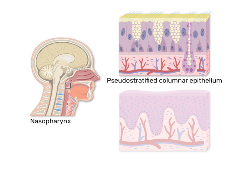
- Nature of epithelium: Respiratory epithelium (ciliated, columnar, and pseudostratified)
- Causes of obstruction: polyps, mucus
- Pharyngeal opening of the auditory tubes: It is the site where the auditory tube (the tube connecting the middle ear and pharynx) opens into the nasopharynx.
- Both superior and anterior aspects are innervated by the maxillary nerve.

Figure 3: An important point to note is that the nasopharynx has “pseudostratified columnar epithelium”. Image Credit: GetBodySmart.
Oropharynx
- Oropharynx is defined as the portion behind the oral cavity. It extends from the uvula up to the hyoid bone.
- Anterior opening: into the mouth
- Inclusions:
- Palatine tonsil (in the lateral wall)
- Tonsillar fossa (in the lateral wall)
- Tonsillar (faucial) pillars (in the lateral wall)
- The base of the tongue (in the anterior wall)
- Epiglottic vallecula (in the anterior wall)
- The inferior surface of the soft palate (in the superior wall)
- Uvula (in the superior wall)
- Epiglottis (closes over the glottis after we swallow the food; prevents aspiration)
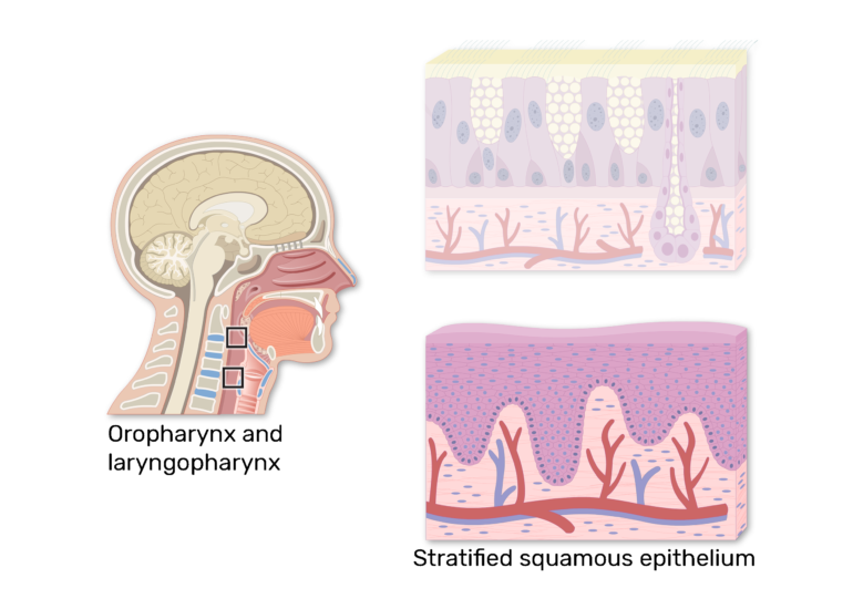
- Nature of epithelium: Non-keratinized and squamous stratified
- Organisms living in the oropharynx region: HACEK organisms
- H stands for Haemophilus
- A stands for Actinobacillus actinomycetemcomitans
- C stands for Cardiobacterium hominis
- E stands for Eikenella corrodens
- K stands for Kingella
Laryngopharynx
- Laryngopharynx is defined as the caudal part (part connecting the throat and esophagus) of the pharynx.
- Position: Inferior to the epiglottis
- 2 pathways of laryngopharynx:
- Respiratory (laryngeal) pathway
- Digestive (esophageal) pathway
- Functions: Conduit for food and liquid to the stomach
- 3 major sites of the laryngopharynx:
- Pyriform sinus
- Postcricoid area
- Posterior pharyngeal wall
- Nature of epithelium: Stratified squamous epithelium
- Inclusions:
- Vascular supply:
- Superior thyroid artery
- Lingual artery
- Ascending pharyngeal artery
- Neural supply:
- Vagus nerves (plus Arnold’s nerve)
- Glossopharyngeal nerve
- Innervated by:
- Pharyngeal plexus (connects the pharynx and superior cervical ganglion)
- Recurrent laryngeal nerve
- Vascular supply:
Figure 4: An important point to note is that in contrast to the nasopharynx, both the oropharynx and laryngopharynx have “non-keratinized squamous stratified epithelium”. Image Credit: GetBodySmart.
Muscular Components Of The Pharynx
There are two major muscular components of the pharynx.
- Circular constrictor muscles: Forms the outermost layer and function as constrictors (tightening of the pharynx). It is innervated by the vagus nerve. The four main muscles in the exterior wall of the pharynx are:
-
- Superior pharyngeal constrictor (superior constrictor muscle)- It originates from, the pterygoid hamulus, a medial pterygoid plate of the sphenoid bone, mylohyoid line of the mandible and pterygomandibular ligament) and inserts into the occipital bone’s pharyngeal tubercle and the pharyngeal raphe (midline fibers) posteriorly. This allows the closure of the soft palate while the swallowing process and propels bolus downwards)
- Middle pharyngeal constrictor
- Inferior pharyngeal constrictor
- Stylopharyngeus

Figure 5: Different muscles in the pharynx. (Superior pharyngeal constrictor, middle pharyngeal constrictor and inferior pharyngeal constrictor muscles). Image Credit: StatPearls Publishing LLC.. -
- Inner longitudinal muscles: Lies underneath the outer circular constrictor muscle layer. This has 2 muscle bands. 2 of the 3 inner muscle layers are innervated by the vagus nerve.
NOTE:
- Blood supply of muscles: Pharynx’s muscles get their blood supply from the external carotid arteries. These arteries branch into several smaller arteries. Each of these smaller arteries feeds a unique muscle area in the pharynx. The blood ultimately exits from the pharyngeal plexus (network of veins) into the internal jugular vein (IJV). These IJVs run along our necks.
- The temporal bone is connected to the pharynx by the muscles of the pharynx.
Pharynx Function
Pharynx serves roles in both the digestive and respiratory systems.
Digestive: The food ingested via mouth travels through the pharynx to the food pipe (esophagus) to finally reach the stomach.
Respiratory: The air inhaled via the nostrils (and mouth) travels through the pharynx, larynx and windpipe (trachea) to finally reach the lungs.
Other roles:
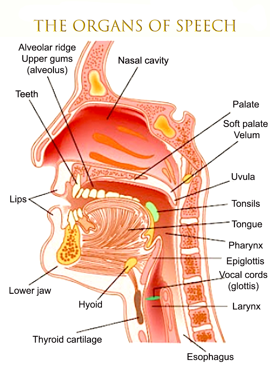
- In human speech
- As resonating chamber (phonation)

Figure 6: Pharynx plays an important and lesser-known role in speech. Image Credit: World Language Gazetteer –
Clinical Significance
Pharynx has a lot of clinical significance associated with it.
-
Inflammation
Pharyngitis is a medical term used to describe the inflammation of the pharynx (sore throat).

Figure 7: There could be different causes for pharyngitis. Image Credit: The College of Family Physicians of Canada-
-
Dysphagia
Dysphagia is a medical term used to describe the condition of acute difficulty or discomfort in swallowing food/liquids. It can occur due to a variety of reasons, including muscle weakness in the pharynx and other parts of the throat.

Figure 8: There are different causes of dysphagia, one being a weakness in the pharyngeal muscles. Image Credit: VeryWell.
-
Pharyngeal Cancer
Pharyngeal cancer is the medical term used to describe the cancer originating in the neck/throat.
-
Sleep Apnea
Some studies have shown a correlation between sleep apnea (a sleeping disorder) and some abnormalities in the pharynx. When the muscles at the back of your pharynx (throat) don’t relax properly, it may lead to discomfort in breathing as you sleep.
-
Pharyngeal venous plexus and bleeding
The pharyngeal venous plexus can be a source of bleeding in certain situations. For example, trauma to the head and neck region can pose risks to the veins in the pharyngeal venous plexus. This could rupture the veins leading to bleeding and potential complications.
-
Waldeyer’S Tonsillar Ring
Waldeyer’s tonsillar ring is the medical term used to describe a ring of lymphoid tissues located in the larynx. Some important ones are adenoids and tonsils (tubal, palatine and lingual tonsils). Waldeyer’s tonsillar ring is also commonly referred to as Nasal-Associated Lymphoid Tissue (NALT).

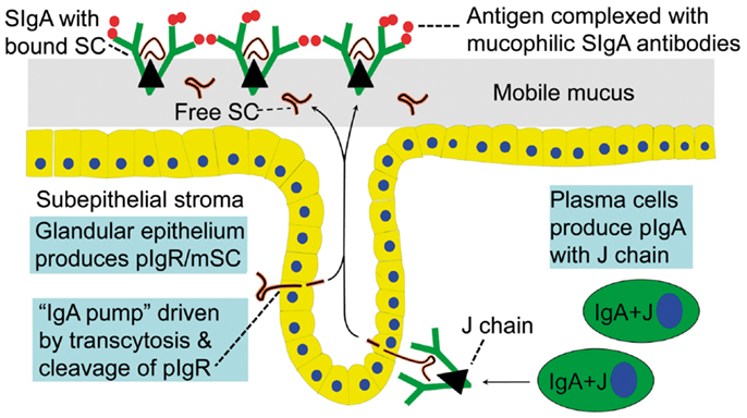
Figure 11: Structure and mechanism of action of secretory immunoglobulin A (sIgA). Image Credit: Per Brandtzaeg.
FEATURED!
“Pharynx and Immunity”
The human body is a natural marvel and the way it is meticulously designed is awe-inspiring. The lymphoid tissues of Waldeyer’s tonsillar ring are located at the gateway of the respiratory and alimentary canals. Since these tonsils are the first site where the human body’s internal system is exposed to air, water, food, and all the microorganisms accompanying these elements, they (tonsils) should be properly geared to counter microbial offenses. Shouldn’t they be?
So, this is the reason that tonsils are well-equipped with NALT and MALT. While NALT stands for Nasal-Associated Lymphoid Tissue, MALT stands for Mucosa-Associated Lymphoid Tissue. Both of them serve as the “first line of defense” against any microbial offense or exogenous aggressors.

Figure 10: Mucosa- and Skin-Associated Lymphoid Tissues. Image Credit: Tak W. Mak.
For this reason, the functions and roles served by the tonsils and other parts of the pharynx are of great significance. Some of the most notable features are:
- The presence of important immune cells (lymphocytes, macrophages, and dendritic cells) in the MALT ensures that pathogens and invaders are detected on priority and destroyed without any delay.
- Production of secretory immunoglobulin A (sIgA) in the pharynx ensures that the mucosal surfaces and mucous membranes of the body can effectively prevent bacterial and viral infections.
- Pharyngeal microbiota (community of healthy microorganisms) ensure the maintenance of a healthy immune system as they help in training the immune cells and actively compete with harmful pathogens for resources, thus minimizing their chances of survival.
So, the next time you swallow or speak, take a moment to appreciate the amazing immune function of your pharynx!
Figure 11: Structure and mechanism of action of secretory immunoglobulin A (sIgA). Image Credit: Per Brandtzaeg.
Take the

Further Reading
References
- Malhotra, A., & White, D. P. (2002). Obstructive sleep apnoea. The lancet, 360(9328), 237-245.
- Hellings, P., Jorissen, M., & Ceuppens, J. L. (2000). The Waldeyer’s ring. Acta oto-rhino-laryngologica Belgica, 54(3), 237–241.
- Fasick J (2006). Respiratory Syster. Benjamin Cummings (Pearson Education, Inc). p. 1.
- Graham A, Richardson J (October 2012). Developmental and evolutionary origins of the pharyngeal apparatus. EvoDevo. 3 (1): 24. doi:10.1186/2041-9139-3-24. PMC 3564725. PMID 23020903
- Elhakeem, A.A. (2021). Adenoid and Tonsils. In: Al-Qahtani, A., Haidar, H., Larem, A. (eds) Textbook of Clinical Otolaryngology. Springer, Cham. https://doi.org/10.1007/978-3-030-54088-3_56
- Mak, T. W., & Saunders, M. E. (2006). Cells and Tissues of the Immune Response. The Immune Response, 35–67. doi: https://doi.org/10.1016/B978-012088451-3.50005-3
- Brandtzaeg, P. (2013). Secretory IgA: designed for anti-microbial defense. Frontiers in immunology, 4, 222.
©BiologyOnline.com. Content provided and moderated by Biology Online Editors.


:max_bytes(150000):strip_icc()/esophageal-dysphagia-5097624-Color-v2-d3736ff073c04c2385ed0d5909ac969e.jpg)