
Cytology
n., plural: cytologies
[saɪˈtɑləd͡ʒi]
Definition: the study of cells
Table of Contents
The biological world is so diverse that studying it under a single umbrella as a subject is next to impossible. Hence, scientists prefer to distinguish between different specialized fields and study them in depth separately. There are many different fields in basic and advanced life sciences like biochemistry, biophysics, physiology, anatomy, genetics, molecular biology, histology, botany, zoology, and so on. Cytology is one of these various fields. Cytology is the study of biological cells. Since all living organisms are made up of the basic life-forming unit called ‘cell’, hence cytology/cell study gains attention whether it be plant or fungal system, animal system, bacterial system, or viruses. A person who studies cell biology is commonly referred to as a cytologist or cytopathologist. Since cytology has been long referred to as “cell biology”, cytologists are also referred to as cell biologists. Let’s move on and learn about what all cytology encompasses, how we define cytology in different contexts, and much more.
Cytology Definition
What is cytology? Cytology as a subject can be defined in different ways depending upon the context of discussion like anatomical, medical, in life science (general biology), etc. Let’s take up each one of them here.
Biology Definition
Anatomy Definition
In Anatomy, cytology is defined as the study of cells for making a diagnosis based on the abnormalities encountered in the anatomical structures of the human body and then correlated to different types of cells. This serves as a deciding ground for making a diagnosis for a patient.
Medical Definition
In the medical field, ‘cytology’ refers to a type of medical investigation of a specific type of cell. Most commonly, this singular type of cell is extracted to investigate and make a medical diagnosis for patients. Medical practitioners recommend these cytologic tests to look into changes, abruptions, and alterations typical to some disease, disorder, or malfunction. Such changes at the cellular level demand cell extraction from patients in the form of fluid specimens (blood, urine, respiratory phlegm, etc), swabs (e.g., vaginal, cheek, and nasal), biopsies, etc. These medical cytology tests (cytopathological tests) help doctors and medical practitioners in narrowing down their dimensions of diagnosis for the illness. Sometimes these tests also aid in the early detection of cancers, tumors, abnormalities, detection of infectious organisms, congenital diseases, and much more.
Watch this vid about cytology
NOTE: People often confuse between “cytology” and “histology”
- Medical cytology exams usually refer to investigations of a single type of cell in the human body.
- Medical histology exams usually refer to investigations of an entire block of tissue from the human body.
Table 1: The basic difference between cytology and histology. | |
|---|---|
| Cytology | Histology |
| Pertains to “single cell type” | Pertains to “tissues” (A tissue is a group of cells) |
| This research area is very narrow and concentrated one | This research area has a wider approach |
| Sample processing: simple and easy | Sample processing: complex and a little difficult for new hands |
| Focuses on different aspects of cell biology, .g., cell behavior, communication, cell-to-cell interaction, biochemistry, etc. | Focuses on mainly tissue architecture, meaning how it is made, what it is composed of, etc. |
Data Source Akanksha Saxena of Biology Online.
Cytology Examples
Some examples of cytology examples from a medical perspective are:
- Ear Cytology
- Breast Cytology
- Mesothelioma Cytology
- Macrophage Cytology
- Veterinary Cytology: Cytology of animal cells.
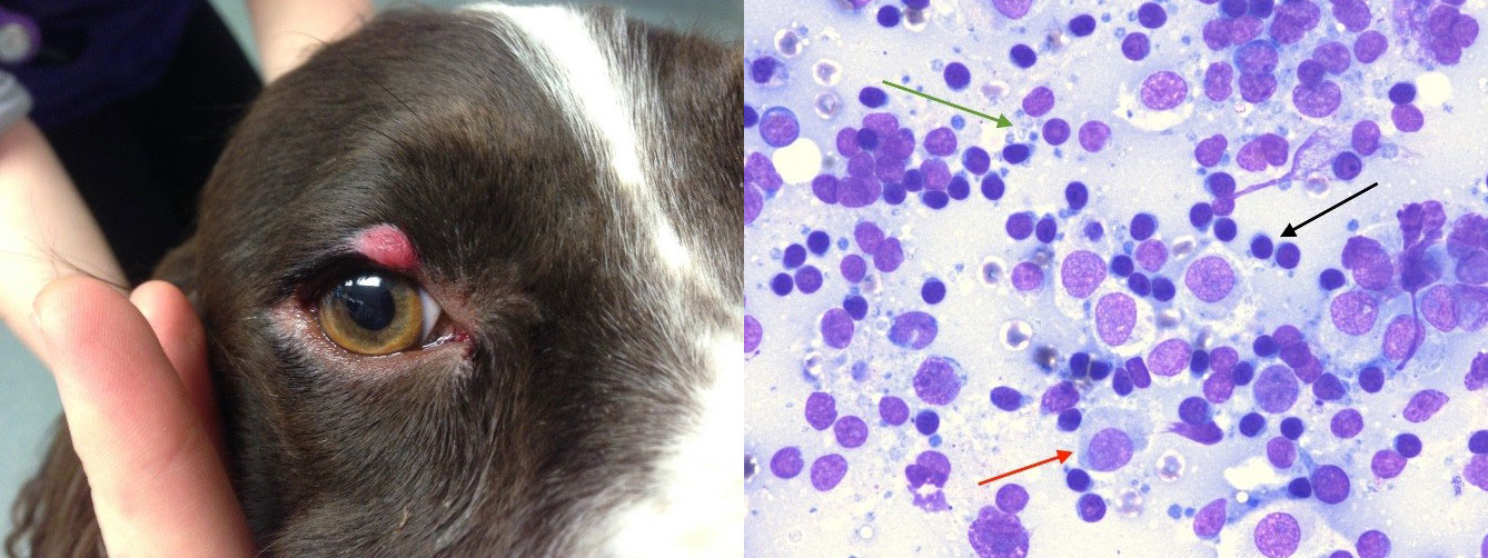
- Histiocytoma Cytology: A histiocytoma is an abnormal development of a cellular mass in the immune cell. This is common to both dogs and the human immune system. When occurring in dogs, this is usually a benign one (not infectious and dangerous). But when happening to humans, this leads to a disease development namely, Hashimoto-Pritzker disease.
- Non-Gyn Cytology: The use of cytology in diagnosing medical conditions in body tissues other than gynecological (or female body) specific manner is called non-gyn cytology.

Cytology Types
When we talk about cytology in the light of medical science, we usually categorize it into two groups. The two types of cytology are exfoliative and intervention cytology. Some pointers common to both of these categories are:
- You can study any type of body cell in cytopathology for learning about the cell’s nature, metabolism, cell-to-cell interaction and communication, biochemistry and cell physiology, cell composition, and cell cycle.
- Since human medical cytology encompasses studies of human body cells at the “microscopic level”, only a small amount of sample is enough for this study whether it be the study of epidermal cells, blood cells, tumor cells, or any other type of cells.
Exfoliative Cytology
Exfoliative cytology is a subset of cytology where medical practitioners extract cells for cytological studies from the “natural shedding” or “surface scraping” i.e., exfoliation of the body or body organs. This is a non-invasive way to collect cell samples. The practitioner will at maximum, only scrape or brush or exfoliate the body’s or organ’s surface for the tissue collection. Some examples of exfoliative cytology are:
1. “Natural shedding” way of cell sample collection
- From spits, mucus, phlegm, nasal vapors, sputum. (For respiratory cytology test)
- From urine (For urine cytology test). This test for the detection of atypical urothelial cells (AUC) can help in the early detection of urinary tract carcinoma.

- From discharges of the body like tears, vaginal discharge, penis discharge, semen, glands discharge, discharge from nipples (For fluid cytology test)

2. “Surface scraping” way of cell sample collection
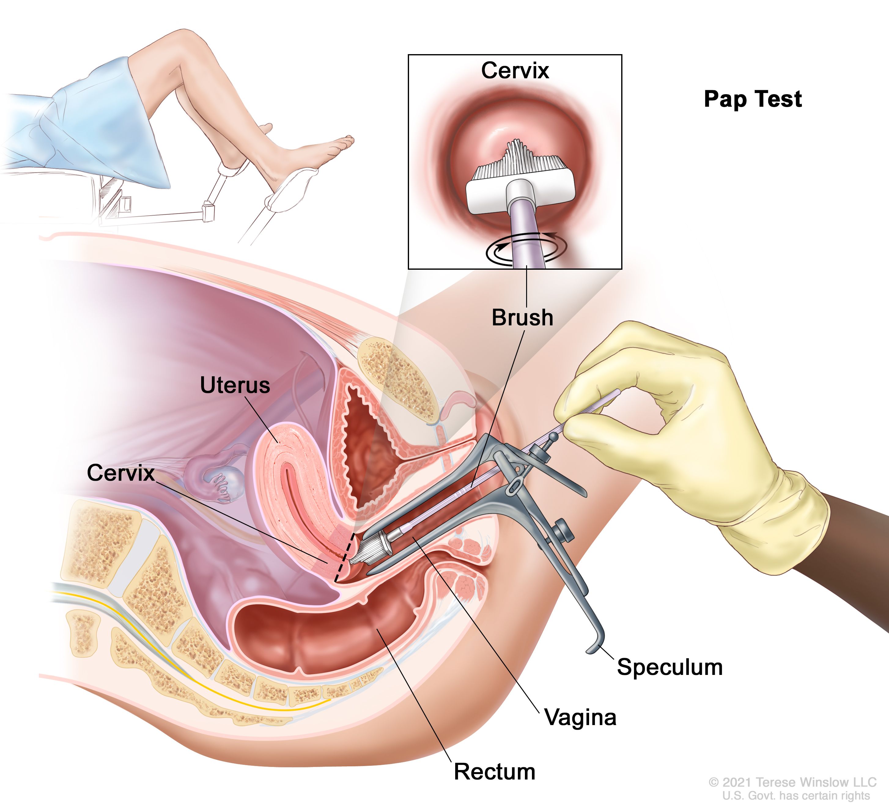
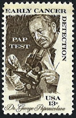
- Exfoliation or scraping of the cells from a woman’s cervix i.e., a pap smear, is a type of gynecologic cytology test. This is commonly performed to check for cervical cancer or to trace any cellular level changes that can indicate the onset or probability of cervical cancer (also called cervical cytology examination). This is also performed for conditions like inflammation, serious infections in female genitalia, for certain types of HPV (human papilloma virus). Pap smears are also called the Papanicolaou test.

- Exfoliation or scraping of the cells from the gastrointestinal tract is a type of exfoliative cytology test in which a healthcare practitioner will brush off some cells from the gastrointestinal tract lining. This can be from the lining of the stomach or intestines, etc using an endoscopy process.

(A- surface epithelial cells, B- Goblet cells, C- adenocarcinoma cells, D- Distinctly-enlarged adenocarcinoma cells from a gastric ulcer, E- Irregular shape of the nucleus in these adenocarcinoma cells are a critical finding for making a diagnosis here, F- cell sarcoma cells, G- lymphoblast cells, H- Multinucleated huge cells showing Hodgkin’s disorder of the stomach, I Benign cells from gastric ulcers.) Image Credit: Raskin, H.F., 1960.
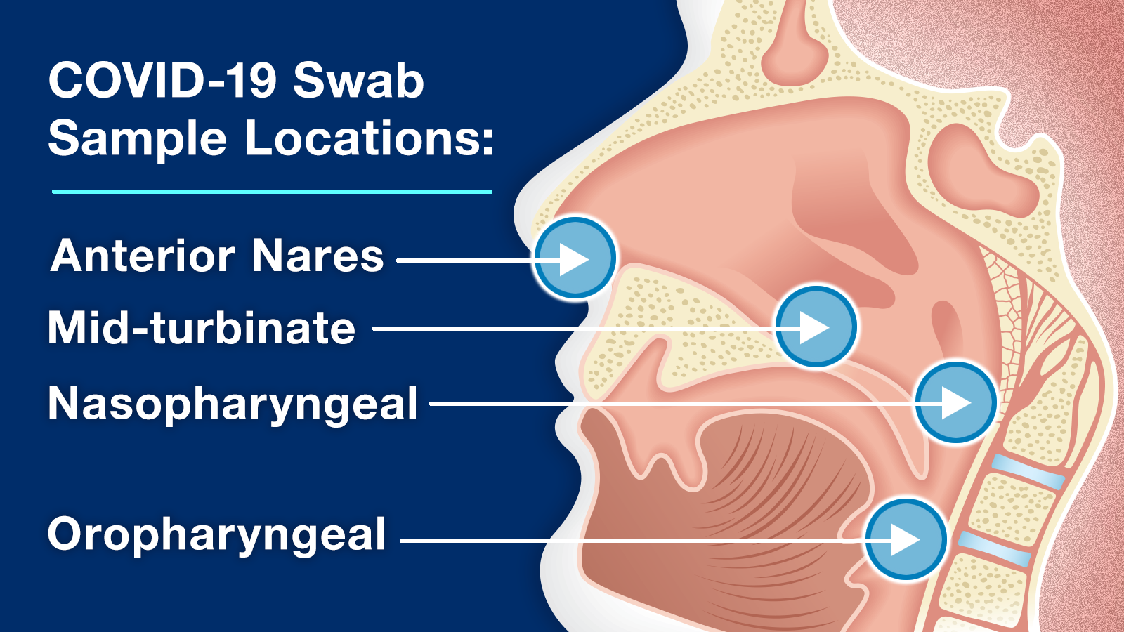
- Exfoliation or scraping of the cells from the skin surface (skin cytology test) or mucous membrane surfaces of the nose, throat, or mouth are also very common. This is the test done for the collection of samples for RT-PCR for SAR-CoV-2 (Covid Virus) detection.

Intervention Cytology
Intervention cytology is a subset of cytology where medical practitioners extract cells for cytological studies by “invasive or intervening process” with your body. In order to access cell samples for some medical examinations, such invasive procedures are unavoidable. For this, your doctor might have to pierce a hole to make their way inside the concerned organ. There are as such no defined categories for this type of cytology testing.
FNA Cytology Test
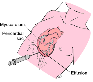
FNA stands for fine-needle aspiration. This is one of the most common cytology testings. In this, a very thin, fine needle is pierced into the area from where your healthcare provider requires a cell sample. Some examples where an FNA is usually performed are: for drawing samples for fluid-filled cysts under a patient’s skin., for lymph nodes, for pleural fluid cytology (fluid in the spaces between the two lungs and inside chest wall), for some suspicious solid lumps, globules, nodules, fatty balls or collection of masses under the skin in any part of the body or even for pericardial fluid cytology (fluid in heart sacs)

Interesting Fact
He first observed smears of cervical cells on slides in the 1917 and 1928 timelines. Using this test, one can detect early precursor lesions on upcoming cervical cancer development in a lady. As the test gained recognition and popularity, more and more women across the globe started getting tested. This tremendously helped in the early detection of cervical cancer cases in the mid-1900s. According to an estimate, the death toll taken by cervical cancer alleviated sharply after the 1960s.
Such an invention in the field of medical biology won’t have been possible without the study of cytology! 🙂

Try to answer the quiz below to check what you have learned so far about cytology.

References
- Raskin, H.F., Pletieka, S. (1960). Exfoliative Cytology of the Stomach. Cytology Series. Page 82-89. https://acsjournals.onlinelibrary.wiley.com/doi/pdfdirect/10.3322/canjclin.10.3.82
- Polaski, A., Tatro, S.E. (1996) Luckmann’s Core Principles and Practice of Medical-surgical Nursing. Saunders. 1776 pages. ISBN0721659942 (ISBN13: 9780721659947)
- Brimo, F., & Auger, M. (2016). The atypical urothelial cell category in the Paris System: Strengthening the Achilles’ heel. Cancer cytopathology, 124(5), 305–306. https://doi.org/10.1002/cncy.21668
- Barkan G.A. et al. (2016) Atypical Urothelial Cells (AUC). In: Rosenthal D., Wojcik E., Kurtycz D. (eds) The Paris System for Reporting Urinary Cytology. Springer, Cham. https://doi.org/10.1007/978-3-319-22864-8_4
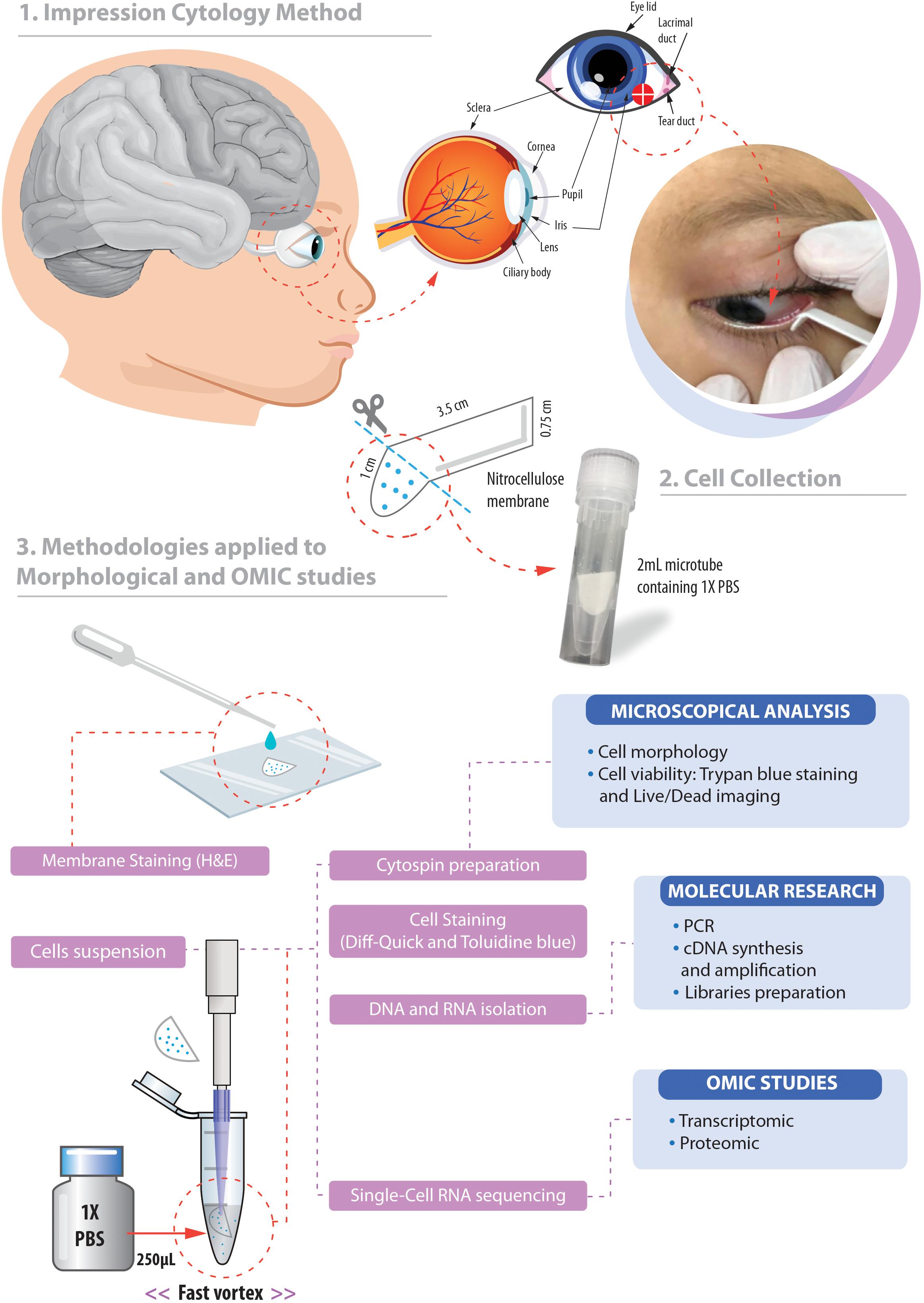
- Barbosa, R. H., Dos Santos, M., Silva, T. P., Rosa-Fernandes, L., Pinto, A., Spínola, P. S., Bonvicino, C. R., Fernandes, P. V., Lucena, E., Palmisano, G., Melo, R., Cardoso, C., & Lemos, B. (2019). Impression Cytology Is a Non-invasive and Effective Method for Ocular Cell Retrieval of Zika Infected Babies: Perspectives in OMIC Studies. Frontiers in molecular neuroscience, 12, 279. https://doi.org/10.3389/fnmol.2019.00279
- Al-Abbadi M. A. (2011). Basics of cytology. Avicenna journal of medicine, 1(1), 18–28. https://doi.org/10.4103/2231-0770.83719
©BiologyOnline.com. Content provided and moderated by Biology Online Editors.


