
Peritoneum
n., plural: peritoneums or peritonea
[ˌpɛɹ.ɪt.əˈniː.əm]
Definition:The membrane lining of the peritoneal cavity or coelomic cavity
Table of Contents
What is the Peritoneum?
The term peritoneum refers to the serous membrane that constitutes the biologically active inner lining of the abdominal cavity and the pelvis in human beings. It constitutes both the parietal layer (lines abdomen and pelvis) and the visceral layer (surrounds abdominal organs). Peritoneum also refers to the lining of the coelom (the main body cavity in all members of the Kingdom Animalia) in amniotes. Only some members of the invertebrates have peritoneum like Annelids. The nature of the peritoneum is “mesothelial”, i.e. it is composed of simple squamous epithelial cells with a “mesodermal origin” with some support derived from a thin layer of fibrous connective tissue underneath.
As emphasized in the Embryology, Fetus, and Blastula lessons, the peritoneum is derived from the “embryonic mesoderm cell layer” which comes into existence from the “inner cell mass” of blastula during embryonic and fetal development. You can engage in more detailed descriptions of embryonic and fetal development here: Embryology, Fetus, and Blastula.
Since the peritoneum plays a very essential role in encapsulating all major abdominal organs in the human body while also serving as a channel for the organ’s nerve connections, blood supply, and lymphatic vessels, it becomes imperative to study its intricate structure, location, development, and clinical significance. Read on to learn more about all of these aspects.

Watch this vid about the peritoneum:
The peritoneum refers to the serous membrane that constitutes the biologically active inner lining of the abdominopelvic cavity or coelomic cavity. In humans, it lines the abdomen and is folded over the viscera. It is made up primarily of mesothelium. There are two layers: the parietal peritoneum (the outer layer that lines the abdomen and the pelvis) and the visceral peritoneum (the inner layer that surrounds abdominal organs). In between these two layers is a space filled with serous fluid. This space is referred to as the peritoneal cavity. In other animals, the peritoneum lines the coelom cavity. Its functions are to support the abdominal or coelomic organs and to serve as a conduit for their blood vessels, lymph vessels, and nerves.Etymology: Ancient Greek perí (“around”) + tónos (“stretch”) + –aios (“suffix to form an adjective”)
Where is the Peritoneum Located?
Peritoneum is inclusive of both the inner lining of the abdominopelvic cavity and the outer covering of visceral organs (both abdominal and pelvic organs). This is the reason that we identify the location of the peritoneum to be both in the abdominal cavity and pelvic cavity while also including a wall of organs.

What is the Function of the Peritoneum?
Peritoneum serves several essential functions in the human body. The same are listed below.
- Provision of padding and insulation to the abdominal organs. (due to the presence of peritoneal fats in the layer of the peritoneum)
- Ensuring that organs are held in their proper place within the abdominopelvic cavity. (due to the presence of peritoneal ligaments which attach organs amongst themselves and to the back of the abdomen’s wall)
- Prevention of friction between different organs on movement by providing lubrication. (due to the presence of peritoneal fluid)
- Provision of immunity to the peritoneal cavity (due to the presence of the ability to filter pathogens and heal itself quickly)
- Serving as a channel for circulatory, nervous, and lymphatic systems that support vital abdominal organs. (due to the presence of blood vessels, nerves, and lymphatic vessels)

Structure
The structure of the peritoneum is understood as a “serum-secreting membrane”, hence called the serous membrane. Such a membranous structure is present in other human body cavities too and is commonly referred to as the mesothelium.
The mesothelial nature of the membrane stands for a top epithelial cell layer accompanied by a lower connective tissue (fibrous tissue layer).
Roles of different tissue types in the peritoneum:
-
- Top epithelial cell layer: It plays an essential role in the secretion and absorption of fluids. It also helps in the filtration of unwanted particles. Also, it helps as a conduit for blood, lymph, and nerve supply.
- Bottom connective tissue layer: It plays an integral role in holding everything together. It has been reported that this layer also helps in anchoring and attachment of some organs to the abdominal wall while ensuring the suspension of others inside the cavity.

-
Layers
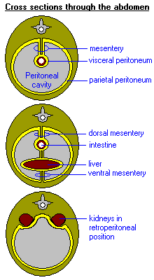
- There are two essential layers in the peritoneum. The outer layer that forms the lining of the abdominopelvic cavity is called the parietal peritoneum and the inner layer that covers the visceral organs is called the visceral peritoneum. Within the two layers lies the peritoneal cavity — a potential space.
- The parietal peritoneum is thicker than the visceral peritoneum.
- One of the vital roles of the parietal peritoneum is the formation of the serous membrane of the testes in males. The parietal peritoneum forms the “tunica vaginalis” by the outpouching (vaginal) process (Figure5)
- One of the vital roles of the visceral peritoneum is the formation of the mesentery in the GIT (gastrointestinal tract). The mesentery is formed by two layers of visceral peritoneum. It helps in the attachment of nerves and vasculature (blood vessels and lymphatic network) to the organs lying in the peritoneal cavity.

-
Subdivisions
The peritoneum is further subdivided into several regions based on its function and location. We discuss the six important subdivisions of the peritoneum here. To begin with, the peritoneal folds include omentums (or omenta), mesenteries, and ligaments. These folds function to connect organs or to the abdominal wall.
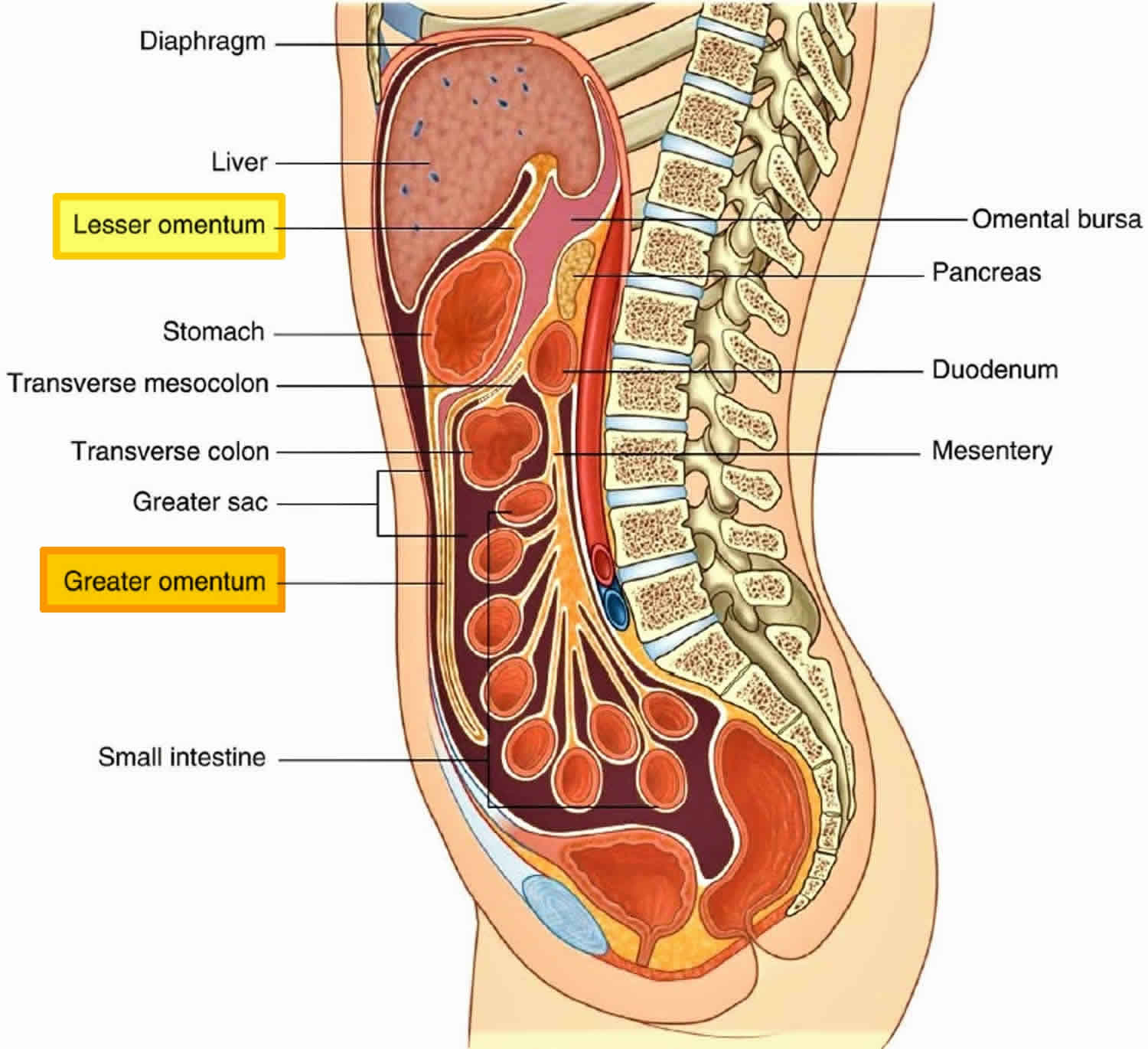
The peritoneal cavity is divided into two main regions — (1) the greater sac and (2) the lesser sac, which are connected by the omental foramen. The lesser sac is further subdivided into two omenta — (1) greater omentum and (2) lesser omentum.
- Greater omentum: The greater omentum is a fatty sheet that hangs like an apron from the greater curvature of the stomach and extends down over the intestines. It plays a crucial role in the immune system by trapping and isolating infections and toxins.
- Lesser omentum: The lesser omentum is a double layer of peritoneum that extends from the liver to the lesser curvature of the stomach and the beginning of the duodenum. It contains the “portal triad”, which includes the portal vein, hepatic artery, and common bile duct.
- Mesentery: The mesentery is a fan-shaped fold of the peritoneum that connects the small intestine to the posterior abdominal wall. It contains blood vessels, lymphatic vessels, and nerves that supply the small intestine.
- Mesocolon: The mesocolon is a broad fold of the peritoneum that attaches the colon to the posterior abdominal wall. It helps to anchor the colon in place and provides blood vessels and lymphatic vessels to the colon.
- Falciform ligament: The falciform ligament is a thin sheet of peritoneum that attaches the liver to the anterior abdominal wall and diaphragm. It contains the ligamentum teres, which is a remnant of the umbilical vein.
- Retroperitoneum: The retroperitoneum is the space behind the peritoneum that contains the kidneys, adrenal glands, pancreas, and parts of the duodenum and colon. These organs are covered by the parietal peritoneum on their anterior surface only.

-
-
Omenta
-
Table 1: Detailed description of different omenta of the lesser sac of the peritoneal cavity. | ||||
|---|---|---|---|---|
| Omentum | Description | Function | Ligaments | Location |
| Lesser omentum | A double layer of peritoneum that extends from the liver to the lesser curvature of the stomach and the beginning of the duodenum | Provides support and stability to the stomach and duodenum | Hepatogastric ligament, hepatoduodenal ligament (same autonomic nerve supply) | Between the liver and the stomach/duodenum |
| Greater omentum | A fatty sheet that hangs from the greater curvature of the stomach and covers the intestines before curving back upwards to attach to the transverse colon | Provides insulation, protection, and immune support to the abdominal organs | Gastrocolic ligament, gastrosplenic ligament, Gastrophrenic ligament, Splenorenal ligament | In front of the small intestines and transverse colon |
Data source: Dr. Harpreet Narang of Biology Online
-
-
Mesenteries
-
- Dorsal mesentery: The dorsal mesentery consists of two layers of peritoneum that span from the posterior abdominal wall to the gastrointestinal tract organs.
- Ventral mesentery: The ventral mesentery is a single layer of the peritoneum that extends from the liver to the ventral body wall.
Table 2: Detailed description of different mesenteries of the peritoneum. | |||
|---|---|---|---|
| Mesentery | Number of Layers | Structure | Function |
| Dorsal mesentery | 2 | Contains blood vessels, lymphatics, and nerves that supply the organs it supports. | Provides support and stabilization to the organs of the gastrointestinal tract. |
| Ventral mesentery | 1 | Contains the falciform ligament, which attaches the liver to the ventral body wall. | Provides support and stabilization to the liver. |
Data source: Dr. Harpreet Narang of Biology Online
-
-
Other ligaments and folds
-
There are several ligaments in the peritoneum. To list, some of the important ones are:
» Falciform ligament
» Suspensory ligament of the ovary
» Round ligament of liver
» Coronary ligament
» Round ligament of uterus
» Ligamentum venosum
» Phrenicocolic ligament
» Left triangular ligament
» Right triangular ligament
» Broad ligament of the uterus
There are several folds in the peritoneum. To list, some of the important ones are:
» Umbilical folds
» Ileocecal fold
-
Classification of abdominal structures
Abdominal structures are classified into four major categories.
- Intraperitoneal: Ovaries (in women), stomach, some part of the duodenum (half of the 1st part), jejunum, transverse colon, ileum, cecum, appendix, sigmoid colon, some part of the rectum (upper one-third), spleen and some part of the pancreas (only tail part).
- Mesoperitoneal: Liver
- Retroperitoneal/Extraperitoneal: Some part of the duodenum (remaining), inferior vena cava, ascending and descending colons, adrenal glands, some part of the rectum (middle one-third), proximal ureters, aorta, some part of the pancreas (except the tail), kidneys and renal vessels are primarily retroperitoneal organs.
- Infraperitoneal/Subperitoneal: Some part of the rectum (lower one-third),
gonadal blood vessels, uterus, distal ureters, fallopian tubes, and urinary bladder (males).

-
Development
The development of the peritoneum is crucial for the proper formation and functioning of the digestive organs. Any disruption in this process can lead to developmental abnormalities or malformations that may result in health complications later in life. Understanding the developmental processes that give rise to the peritoneum is essential in studying and treating developmental disorders and peritoneal diseases that affect the digestive system. The developmental biology of the peritoneum is briefly discussed below.
- The peritoneum’s development begins during the embryonic stage.
- It arises from the lateral plate mesoderm.
- This mesodermal layer splits into two layers: the somatic and the splanchnic layers.
- The somatic layer forms the body wall, while the splanchnic layer develops into the viscera and their associated mesenteries.
- During the developmental stages, first, the mesothelium lines the coelomic cavity.
- Next, the mesothelial cells migrate toward the abdominal cavity, and as they do, they secrete a thin layer of connective tissue that eventually becomes the visceral and parietal peritoneum.
- Slowly, as the digestive organs develop, the peritoneum forms the mesenteries.
- Additionally, the peritoneum also forms the omenta.
Clinical Significance
The peritoneum is a crucial structure in the abdominal cavity with substantial clinical significance in various diseases and conditions. Its role in protecting and supporting the abdominal organs highlights the importance of understanding its function and the impact of diseases that affect it.
-
Imaging assessment
Imaging tests are essential in diagnosing and evaluating peritoneum-related problems. The most common imaging tests used to detect peritoneal abnormalities include:
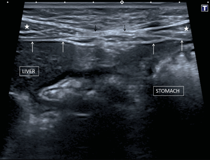
- Ultrasound: This is a non-invasive imaging test that uses high-frequency sound waves to produce images of the internal organs, including the peritoneum. It is a useful tool in detecting fluid accumulation in the peritoneal cavity (ascites) and other abnormalities such as tumors. Although the resolution is less, the amount of exposure to ionizing radiations is also substantially reduced.

Figure 8: Ultrasound of abdomen and peritoneum. Image Credit: James M. Pilcher & Pawan Patel. - Computed Tomography (CT) Scan: CT scans use X-rays to create detailed images of the abdominal organs, including the peritoneum. It is a highly sensitive test that can detect peritoneal abnormalities such as inflammation, infection, and tumors.

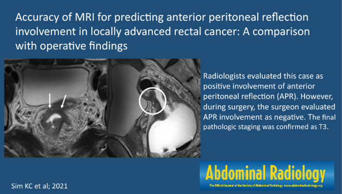
Figure 9: (a) Axial and (b) Coronal CT scans of a person suffering from peritoneal carcinomatosis. One can the large amounts of ascites. Image Credit: Myeong-Jin Kim. - Magnetic Resonance Imaging (MRI): This test uses a strong magnetic field and radio waves to produce detailed images of the body’s internal structures, including the peritoneum. It is a non-invasive test that can provide excellent visualization of the peritoneum and can detect abnormalities such as tumors and inflammatory conditions. The only disadvantage is the long scan time and the issues of motion artifacts. There are some cases where long MRI scans aren’t recommended like peritoneal carcinomatosis, acute pancreatitis, and intraabdominal sepsis.

Figure 10: A study reported high accuracy of MRI for the prediction of anterior peritoneal reflection correlation with rectal cancer. Image Credit: Ki Choon Sim. - X-ray: Although not as sensitive as other imaging tests, an X-ray can detect the presence of gas in the peritoneal cavity, which can indicate the presence of a perforated organ or bowel obstruction.
- Laparoscopy: This is a minimally invasive surgical procedure that involves inserting a small camera into the abdominal cavity to visualize the peritoneum and other abdominal organs. It is a highly accurate test that can detect a wide range of peritoneal abnormalities.
-
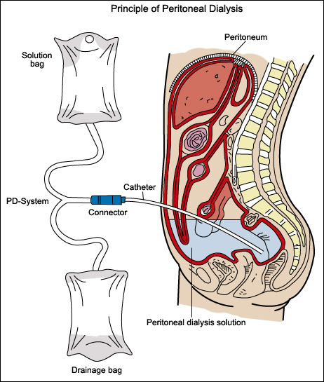
Peritoneal dialysis
Peritoneal dialysis is a method of removing waste products from the blood in the peritoneal cavity. In this intervention, the peritoneal cavity is supplied with a glucose solution through a tube. The glucose solution is left undisturbed in the cavity for a certain period to facilitate the absorption of the waste products. The rich density of vasculature (arteries and veins) in the peritoneal cavity ensures the removal of waste products. Subsequently, the tube is removed.

-
Peritonitis
Peritonitis is a serious condition in which the peritoneum becomes inflamed and infected, usually due to bacterial infection from a ruptured organ, such as the appendix or stomach. Chemical irritation due to gastric/pancreatic juices can also pave the way for peritonitis. It can lead to sepsis, a life-threatening condition that can cause multiple organ failure and death if left untreated.
Common symptoms of peritonitis include:
-
- Fever
- Tenderness
- Pain in the abdominal area (localized or diffuse)
Treatment of peritonitis includes:
-
- Rehydration
- Administration of antibiotics
- Surgical correction of the underlying cause

-
Primary peritoneal carcinoma
Primary peritoneal carcinoma (PPC) is a rare and aggressive type of cancer that originates from the peritoneal lining of the abdominal cavity. It is similar in presentation and treatment to epithelial ovarian cancer, but it differs in the absence of a primary ovarian tumor.
Common presentations:
-
- Abdominal pain
- Bloating
- Ascites
Diagnosis is confirmed through biopsy and imaging tests such as CT scans and MRIs.
Treatment: Combination of surgery, chemotherapy, and radiation therapy. Due to its aggressive nature and late presentation, the prognosis for PPC is generally poor, with a five-year survival rate of less than 30%.
NOTE IT!
Common Dilemmas About Peritoneum!!!
There are some common confusions and dilemmas when the topic of the peritoneum is touched. Let’s shed some light on each one of them and provide a valid explanation for clarity on the topic.
- The confusion between the abdominal cavity and the peritoneal cavity
Explanation: Abdominal cavity is defined as the cavity space of the abdomen. It lies between the abdominal muscles, diaphragm, vertebrae, and pelvic floor. The abdominal cavity is defined by the diaphragm’s muscular wall. On the other hand, the peritoneal cavity lies inside the abdominal cavity (in between the parietal and visceral peritoneum) and is defined by the peritoneum. - Which organs are within the abdominal cavity and peritoneal cavity respectively?
Explanation: Some organs lie within the abdominal cavity but not within the peritoneal cavity. The list of organs in both cavities is provided below.- Organs within the abdominal cavity but outside the peritoneal cavity: These organs are sometimes referred to as “extraperitoneal” in position.
- Aorta
- Esophagus
- Second and third parts of the duodenum
- Ascending and descending colon
- Pancreas
- Kidneys (since they lie behind the peritoneal cavity, kidneys are sometimes specifically referred to as “retroperitoneal” in position.)
- Ureters and bladder (since they lie below the peritoneal cavity, they are sometimes specifically referred to as “subperitoneal or intraperitoneal” in position.)
- Adrenal glands
- Organs within the peritoneal cavity: These organs are sometimes referred to as “intraperitoneal” in position.
- Stomach
- Spleen
- Liver
- First and fourth parts of the duodenum
- Jejunum
- Ileum
- Transverse and sigmoid colons
- Organs within the abdominal cavity but outside the peritoneal cavity: These organs are sometimes referred to as “extraperitoneal” in position.
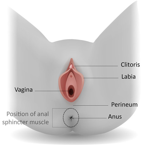
- Are the peritoneum and perineum the same or different?
Explanation: Peritoneum and perineum are not the same parts of the body. While the peritoneum refers to the serous membrane layer of the abdominopelvic cavity, the perineum is the sensitive and small patch of skin between the genitals and anus in the human body. While the peritoneum plays an essential role in the insulation and protection of organs in the abdominal cavity, the perineum plays an important role in processes like defecation, micturition, childbirth, and sexual intercourse. The word peritoneal is used in the context of the peritoneum and the word perineal is used in the context of the perineum.
Figure 13: An illustration to understand the location and structure of the perineum (different from the peritoneum). Image Credit: Royal College of Obstetricians and Gynecologists.
Take the Peritoneum – Biology Quiz!

References
- Alavi, S., Haeri, A., Mahlooji, I., & Dadashzadeh, S. (2020). Tuning the physicochemical characteristics of particle-based carriers for intraperitoneal local chemotherapy. Pharmaceutical Research, 37, 1-24.
- Pilcher, J.M., Patel, P. (2022). Abdominal Ultrasound—Bowel and Peritoneum. In: Walden, A., Campbell, A., Miller, A., Wise, M. (eds) Ultrasound in the Critically Ill. Springer, Cham. https://doi.org/10.1007/978-3-030-71742-1_13
- Sim, K.C., Park, B.J., Kim, M.J. et al. Accuracy of MRI for predicting anterior peritoneal reflection involvement in locally advanced rectal cancer: a comparison with operative findings. Abdom Radiol 47, 508–516 (2022). https://doi.org/10.1007/s00261-021-03356-6
©BiologyOnline.com. Content provided and moderated by Biology Online Editors.


