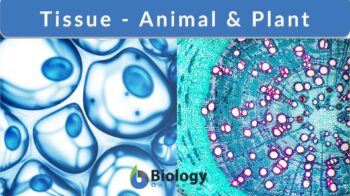
Tissue
n., plural: tissues
[ˈtɪʃu]
Definition: aggregate of cells having the same structure and function
Table of Contents
Tissue Definition
What is tissue in the body? The body tissue is an aggregation of cells that function together and have a similar function. The cells of a multicellular organism do not usually function independently instead they are usually associated with other cells forming a tissue. Mostly, tissues are made up of several types of cells that cover different requirements of tissue function. The study of tissues is called histology. This science studies the anatomy of different tissues using microscopes. The microscopic biological science histology helped in the classification of tissues of both plant and animal tissue as well as identifying their detailed structure.
“When cells of an organism with similar functions and structures come together to form a group, this unit is called tissue.” Get info straight from an Expert: Smooth muscle vs dense regular connective tissue. Join us now!
Types of Animal Tissues
What are tissues? The definition of tissue in animals is a group of cells combined together to perform a certain function. There are four tissue types in animals, each type of tissue has its distinct structure and function. They are (1) epithelial tissues, (2) connective tissues, (3) muscular tissues, and (4) nervous tissues. Each of these tissues has its own unique structure and biological function. These different types of tissues are discussed below:
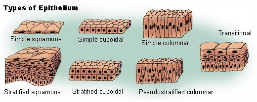
Epithelial tissue
Epithelial tissues (or epithelium) are the most abundant human tissues. They cover the whole surface of the body forming the epidermis layer of the skin. Moreover, it covers the lining of body organs forming mucus lining, such as the digestive system lining. Thus, they protect the organ from external factors, such as injury, pathogenic microorganisms, and loss of fluids. Epithelium contributes to the excretory process where glandular epithelial cells are incorporated in the synthesis and excretion of wastes. Epithelial tissues are also responsible for body secretions and absorption. They are responsible for the secretion of saliva, mucus, sweat, etc. so they form a considerable part of multicellular glands, such as the liver and pancreas. Other epithelial tissue covers the different body cavities to produce secretory lubricants.
Epithelial tissue is either formed of a single layer of epithelial cells (called the simple epithelium) or formed of multiple layers of cells (called the stratified epithelium). Epithelial cells forming a single layer of epithelial tissue have three surfaces: (1) an apical surface, (2) a basal surface, and (3) a lateral surface. Each of these surfaces has a distinct function.
The apical surface of epithelial cells is responsible for exchanging material passing in and out of the cell. The apical surface of epithelial cells of the inner lining of the small intestine contains microvilli that project into the small intestine whereas mucus-moving epithelial cells possess cilia projecting from the apical surface. On the contrary, the surface of the epidermis has no or minimal specialization at the apical surface since these cells are subjected to shedding and abrasion.
The basal surface serves to connect the bottom layer of epithelial tissue to a basement membrane. The basement membrane is a thin non-cellular layer like a sheath. It covers the peripheral nerves and muscles as well as underlying the epithelial tissue. The basement membrane is secreted by different cells. It consists of proteins, such as laminin and collagen. Integrins are proteins in the plasma membranes of epithelial cells that connect them to the basement membranes. In secreting and absorbing cells, the apical surface functions to increase the surface area.
The lateral surface mediates the intracellular connections among epithelial cells. Epithelial cells are connected together forming the epithelial tissues through tight junctions near their apical ends; these junctions do not permit the passage of ions, water, and solutes in the intercellular spaces.
Connective tissue
How does connective tissue differ from the other three major tissue types? Connective tissues contain nonliving extracellular material more than living cells. Relatively few cells are found among connective tissues. They are embedded in the extracellular matrix. The cells are often far from each other except fat tissue cells where adipocytes are packed close to each other.
The main function of connective tissue is to connect different cells and tissues with the aid of some molecules secreted from the connective tissue in the extracellular matrix forming fibrous tissues. Other functions are as follows: providing protection and support to soft tissues, transmit mechanical forces, and production of carbohydrates and proteins.
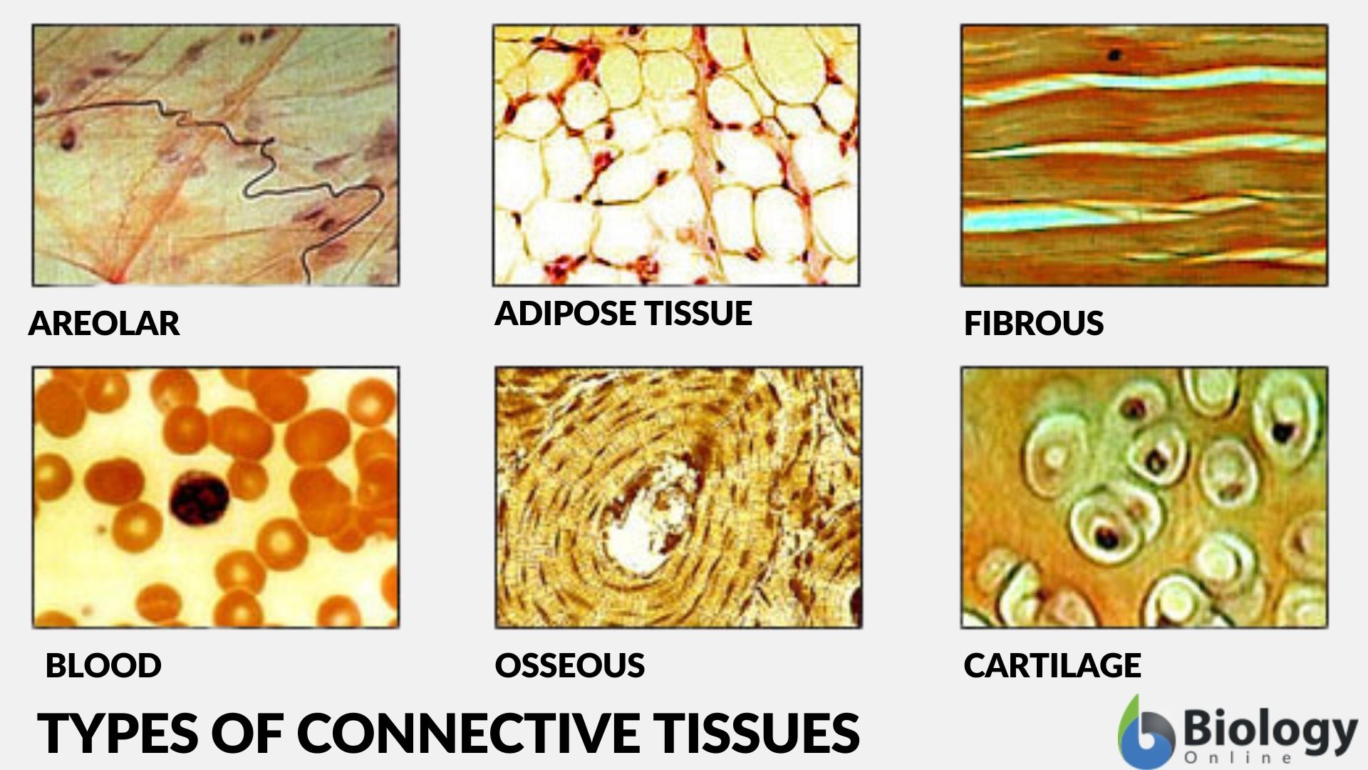
Classification of connective tissues
In humans and other vertebrates, the connective tissues may be classified as follows:
I. Connective tissue proper
A. Dense connective tissue – has more fibers than ground substance.
- Dense regular connective tissue – characterized by collagen fibers that are arranged apparently in one direction, particularly, in a parallel fashion. For example, tendons and ligaments
- Dense irregular connective tissue – characterized by collagen fibers that seem to be arranged in multiple directions. For example, connective tissues found in the lower layers of the skin (dermis) and in the protective white layer of the eyeball
B. Loose connective tissue – has more ground substance than fibers.
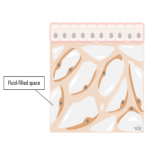
II. Special connective tissue – connective tissues that are not fibrous. Examples: reticular connective tissue, adipose tissue, cartilage, bone, and blood.
Other references classify connective tissues differently; for instance, adipose tissue, a special connective tissue type, is classified as a loose connective tissue (under connective tissue proper).
Connective tissue cells
The cells of connective tissues vary depending on their structure and functions. Fibroblasts, for instance, are a type of connective tissue cells characterized by having abundant endoplasmic reticulum and Golgi apparatus. They produce and secrete fibers, such as collagen, reticular fibers, and elastic fibers. Thus, they are responsible for the maintenance of the extracellular matrix (especially during wound healing or tissue repair). They provide the structural framework for many tissues.
During the embryonic stage, the mesenchymal cells differentiate into different types of connective tissues. Some differentiate into osteoblasts forming bones. Others differentiate into chondroblasts forming cartilages of the deep skull bones.
What type of tissue is blood? Blood is one of the most important connective tissues in the body. It is mainly composed of blood cells and plasma. The blood cells are white blood cells (leukocytes) and red blood cells (erythrocytes). The platelets (thrombocytes) are cellular fragments and so some references do not consider them as blood cells. All of the three types originate from the multipotent stem cells in the red bone marrow. The function of the white blood cells is for protection against infections and for removing cellular debris. The red blood cells carry oxygen throughout the body. The platelets are involved in blood clot formation.
Connective tissue fibers
The fibrous connective tissues are rich in non-cellular components, such as fibers. These fibers are of different types: collagen fiber, elastic fiber, and reticular fiber. The collagen fibers are made up of collagen. They are found in tendons and ligaments. They support the walls of internal organs. They also represent about one-third of the bone weight.
Another type of connective tissue fiber is elastic fibers. They are made up largely of elastin. They are likened to rubber bands due to their elastic property. The elastic fibers can retain their original shape after deformation such as the dermis of the skin, large arteries, and lungs where these tissues containing elastic fibers can stretch and contract in response to different stimuli.
The reticular fibers are fibers made up chiefly of type III collagen secreted by the reticular cells. These fibers are found in different parts of the body, such as the connective tissues supporting fats or lymphoid tissue.
Ground substance
The ground substance is made up chiefly of proteoglycans, glycosaminoglycans, glycoproteins, water, and ions. They serve as space-fillers, absorb shocks, lubricate, and limit the movement of bacteria in connective tissues.
Muscular tissue
Muscles are contractile tissues in the body. The muscular tissue is classified into three types: (1) skeletal muscles that are attached to the bones and help in the body movement, (2) smooth muscles that form the inner lining of blood vessels, and (3) cardiac muscles that form the heart.
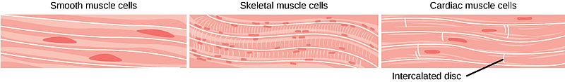
The skeletal muscles are responsible for the voluntary movement of the body. The smooth muscles are for involuntary movement, similar to cardiac muscles. However, the cardiac muscles are similar to skeletal muscles in having conspicuous striations when viewed under the microscope. The cardiac muscles also differ from the other two in having intercalated discs.
READ: The Human Physiology – Muscle
Smooth muscle tissue
The smooth muscles lack the striations when viewed under the microscope. They are made up of spindle-shaped narrow cells with a single, centrally-located nucleus. They lack the characteristic striations because their actin and myosin filaments are not organized in a manner similar to that of skeletal and cardiac muscular tissues. The arrangement of actin and myosin in the smooth muscle cells enables the smooth muscles to contract for a longer time than skeletal muscles with less energy. Therefore, smooth muscles have the ability to maintain the tone of vessels or the uterine contraction during birth. Smooth muscles line the walls of the body tubes such as the digestive system because they are responsible for the movement of material through these tubes. Watch the smooth muscle contraction below.
Smooth muscles as well as the cardiac muscles contract involuntarily, meaning they are not controlled by the conscious demand of an individual. In contrast, skeletal muscles are voluntary muscles that follow conscious control.
Skeletal muscle tissue
Skeletal muscles are multinucleated tissues of fused cells. The muscle cells are fused together forming myotubes. The nuclei of myotubes are large and elongated with a large nucleolus. Skeletal muscle tissues produce great amounts of proteins and RNA. RNA and proteins stimulate the muscle to contract after proteins organize in arrays called sarcomeres. Watch the video below to see how the skeletal muscle contracts.
Skeletal muscles secrete a basal lamina that surrounds the muscle. The basal lamina helps in the transmission of muscle contraction to the surrounding tissues. Satellite cells are located between the basal lamina and the fiber of the muscle. Satellite cells help in the development and regeneration of muscle fibers.
There are two main types of skeletal muscle tissues: fast and slow muscles. They are classified according to their oxygen consumption while contracting. Fast muscles are thicker, stronger, and contract for less period than slow muscles.
Cardiac muscle tissue
Cardiac muscles are striated muscles. They contact like skeletal muscles; however, they are involuntary muscles. Another difference between skeletal and cardiac muscles is that cardiac muscles contain only one nucleus in contrast to multinucleated skeletal muscles.
Cardiac muscles are rich in blood vessels and mitochondria since they need active metabolism to support their continuous contraction. Unlike skeletal muscles, cardiac muscles must contract at the same time to pump the blood from the heart. This effect is achieved by the presence of junctions between cardiac muscles; these junctions link each muscle to the next one so it helps in signal transmission through the entire heart.
Cardiomyocytes (cardiac muscles) do not contain satellite cells as in skeletal muscles; therefore, cardiac muscles do not divide or differentiate. Instead, they grow by increasing their size only.
READ: Heart Anatomy – Biology Online Tutorials
Nervous tissue
Nervous tissues are tissues that form the central nervous system and the peripheral nervous system. Nervous cell tissue forms the brain and the spinal cord in the central nervous system whereas in the peripheral nervous system, they form sensory, motor, and autonomic nerves.
Nervous tissue is formed of large cells called neurons; they have a long body that may reach up to a meter. Neurons have cell processes, dendrites or axons. These cell processes carry signals across different neurons from the cell body to the nerve terminal. In particular, the dendrites carry signals from the periphery to the cell body. The cell body contains numerous organelles that serve its function including ribosomes, cytoplasm, and Golgi apparatus. The axons, on the other hand, contain a large amount of cytoplasm in comparison to the cell body. They also contain microfilaments and microtubules that transport signals and support the cell.
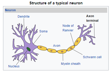
Each axon is branched into regions known as the presynaptic terminals. At presynaptic terminals, the electrical impulse is converted into chemical neurotransmitters that send signals to the following neuron or muscle cell in the chain. Neurotransmitters are stored in pockets and released as a result of the change in the cell membrane permeability when the electrical signal reaches the synapse. Neurotransmitters are released to bind to their receptor on the next cell continuing the transmission of a nerve impulse until it reaches the target tissue. Nervous tissue function is to transmit nerve impulses across different nerve cells in order to control all voluntary and involuntary actions in the body.
The fundamental types of tissues in animals are as follows:
Examples of Plant Tissues
What is tissue in plant biology? We already know that biological tissue is made up of a number of cells performing a specific function. In plants, tissues are similarly made up of cells that perform a particular function. Below are some of the examples of plant tissues that perform a particular function. For example, the plant epidermis is the tissue that renders protection. The ground tissues are the tissues consisting of fundamental plant cells that perform a distinct function. The vascular tissues are the plant tissues involved in the transport of nutrients throughout the plant body. Let’s take a closer look at them below.
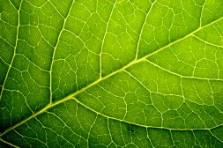
Plant epidermal tissue
The plant epidermal tissue (epidermis) is the most outer protective tissue of the plant. It covers the surface of the plant protecting the inner layers from the outer environment. It is covered by a layer of cuticle that makes the epidermis impermeable to water and therefore helps prevent water loss.
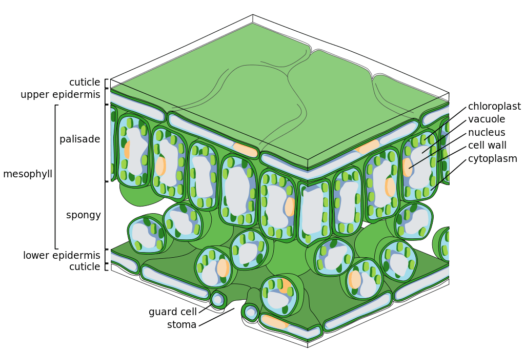

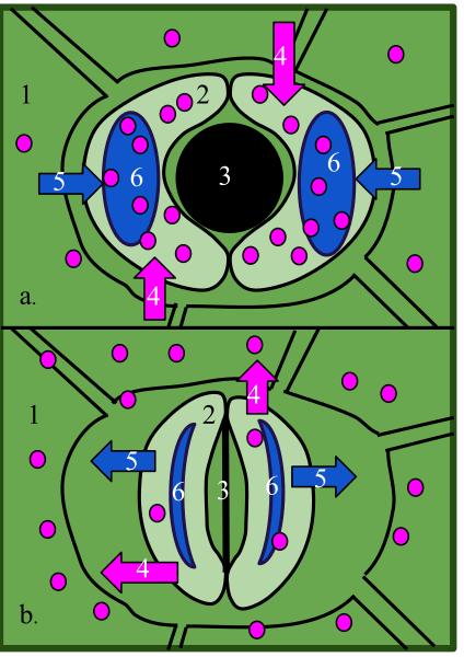
The epidermis of the plant protects it from the ultraviolet radiation of the sun. However, it permits the exchange of gases between the outer environment and the plant via the stomata. The stomata of epidermal tissue are composed of two guard cells. Together, they form the stomatal pore. The inner walls of guard cells are thicker than their outer walls. The guard cells are kidney-shaped. They are attached to each other at their ends with microfibrils of cellulose surrounding them. Guard cells expand when they absorb water, but due to the presence of cellulose microfibrils, they expand longitudinally instead of widely to create a pore that permits the exchange of gases.
Epidermal cells may have extensions called trichomes. Trichomes function to protect the plant from herbivores and insects by secreting toxins or blocking access to the surface of the plant.
Unlike the shoot epidermis, the root epidermis permits the passage of water from the soil to the plant. It is covered by a thinner cuticle that contains shorter waxes. Roots outgrow forming root hairs; these hairs increase the absorbing area of the root (increased intake of water and nutrients). The root epidermis usually produces a hydrophilic carbohydrate that traps water to facilitate its absorption from the soil and to lubricate the area through which the root passes.
Ground tissue
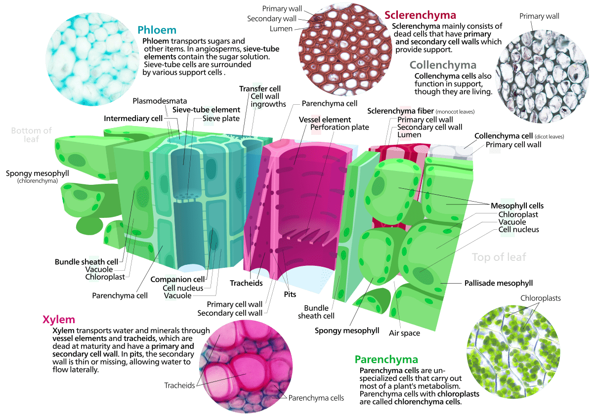
Ground tissues represent all tissues that form the plant which is not epidermal or vascular tissues. Ground tissues are divided into three fundamental plant cell types: parenchyma, collenchyma, and sclerenchyma according to the structure of their cell wall.
The parenchyma cells have a thin wall. They occupy most of the cortex, which is the area between other layers, epidermal, vascular, and pith. Parenchyma stores proteins, starch, and oils in the plant. They also contribute to the growth, development, and support of the plant.
The collenchyma cells are similar to parenchymal cells in having a primary cell wall. The cell wall materials are deposited in the edges of the cell forming collenchyma. Collenchyma cells contribute to the elongation of plant stems. However, they only provide support when the plant is turgid because their cell walls lack hydrophilic substances.
The sclerenchyma cells have thick walls containing lignin. This structure makes sclerenchyma waterproof and strong. The sclerenchyma provides support to the plant more than parenchyma and collenchyma. However, it is more difficult to make thus it is not as abundant as other types of ground tissue. Owing to their rigidity, sclerenchyma may be incorporated in the structure of vessel tissues, it forms the tracheids and vessels in the xylem.
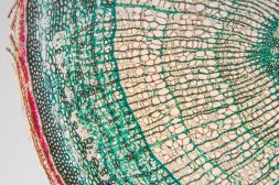
Vascular tissue
The vascular tissues of the plant represent the main system responsible for transportation in the plant, it is composed of xylem and phloem. Vascular tissue is present in all parts of the plant in the form of vascular bundles, stems, leaves, and traversing roots. The xylem transports different materials such as water and ions from roots to leaves and stems of the plant. The phloem transports metabolites, such as amino acids, sugars, and ions produced by the plant from the site of production in leaves to different areas like roots, fruits, and leaves.
The phloem is mainly made up of companion cells, phloem parenchyma cells, sieve tubes, and phloem fibers. Sieve plates separate sieve tubes, these tubes have pores termed as sieve pores. Even though sieve elements lack nuclei, they are living cells containing plasmalemma and proteins. Companion cells surround sieve elements. They contain large nuclei, vacuoles, and dense cytoplasm. Companion cells function to transport metabolites in and out of sieve elements.
The xylem consists of xylem vessels, tracheids, xylem parenchymal cells, and xylem fibers. Large plates join vessels together leaving gaps between vessels, while the ends of tracheids overlap with the following cells with no gaps in between. Lignin is distributed in different forms in vessels such as helical, reticulate, annular, scalariform, and pitted. It can be transported either longitudinally or laterally through pores or pits of plates.
(1) Embryonic or meristematic tissues (e.g. apical meristem and cambium)
(2) Permanent tissues (e.g. epidermis, cork, trichome)
- Fundamental (e.g. parenchyma, collenchyma, sclerenchyma)
- Complex (e.g. phloem and xylem tissues)
(3) Reproductive tissues (i.e. sporogenous tissues).
Tissue Engineering
Tissue engineering is a method of producing tissues outside the body, it combines biological and engineering to repair or create new tissues. This science can affect the medical field greatly by enhancing recovery of damaged tissues due to burns, trauma, or diseases by creating an artificial graft of tissues. Even though this approach seems promising, however, it faces some difficulties such as creating artificial nutritional blood vessels and supplying tissues with circulating blood. Other challenges include manufacturing and using bioreactors to create tissues using the exact right amount of nutrients, enzymes, and cellular components. Cell differentiation in culture might not be controlled so the produced tissue will be hypertrophied and may contain more than one nucleus per cell leading to the formation of a deformed tissue. Therefore, tissue engineering is an emerging promising science that implies the importance and impact of tissue culturing in the field of medicine.
Try to answer the quiz below to check what you have learned so far about tissues.

References
- Bruce M. Carlson (2019). Tissues. The Human Body (pp 27-63). Academic Press. https://doi.org/10.1016/B978-0-12-804254-0.00002-8.
- Enderle, J. (2012). Introduction to biomedical engineering. Academic press.
- Fry, S. C. (2017). Cell walls. Encyclopedia of Applied Plant Sciences,, 1, 161-173.
- Lopez, F. B., & Barclay, G. F. (2017). Plant anatomy and physiology. In Pharmacognosy (pp. 45-60). Academic Press.
©BiologyOnline.com. Content provided and moderated by Biology Online Editors.