
Embryo
n., plural: embryos
[ˈɛmbɹi.əʊ]
Definition: A multicellular organism inthe early stage of development
Table of Contents
An embryo refers to the early developmental stage of a multicellular organism that follows fertilization. This stage represents a crucial juncture in the life cycle, transitioning from a single fertilized cell into a complex organism with specialized tissues and structures. The stage is marked by rapid cell division and the establishment of the basic body plan.

Embryo Definition
In Biology, an embryo is designated as the initial phase of development for a multicellular organism arising after the fusion of a sperm cell (male sex gamete) and an egg cell (female sex gamete released during a woman’s reproductive cycle) during the process of fertilization. This fusion gives rise to a zygote (which is made up of a single cell) which subsequently undergoes a sequence of cell divisions and differentiations ultimately leading to the formation of an embryo.
An embryo is defined as “the stage of development in biological life forms which serves as the precursor to its more intricate and fully developed form as it progresses through subsequent growth and developmental stages”.
Although a term called “pre-embryo” has been doing rounds, this term is not accepted by the scientific community.
The scientific field of embryology delves into “comprehending the mechanisms and phases of embryonic development across diverse organisms”. To read more about this scientific field, head over to our comprehensive article on Embryology where you can learn about comparative embryology, different theories explaining the early embryogenesis process, the science underlying embryonic cleavage, etc.

-
Embryo Etymology
The word embryo is a late Middle English word that is derived from the Medieval Latin word “embrion” which has been further derived from the Greek word “embruon” meaning ‘fetus or little one’. The word embruon can be dissected into two Greek words; “em-” meaning ‘into’ and “bruon” meaning ‘moss’ or ‘something that can swell and increase in size’.
Watch this vid about embryo development:
Biology definition:
An embryo is the stage of early development after fertilization up to the phase of initial organogenesis. It may also pertain to the multicellular organism itself that undergoes extensive and rapid growth and cell differentiation during the early stage of development. In humans, the embryonic stage spans after the zygote phase (at fertilization) to approximately the eighth week from the time of fertilization (note: week eight from the time of fertilization is equivalent to 10 weeks of gestational age).
The timing and duration of the embryonic stage vary widely among species of the animal kingdom — from very few hours in hydrozoan jellyfish to a considerably long embryonic stage in some reptiles, such as the leatherback sea turtle (Dermochelys coriacea), which has an incubation period of around 60 to 70 days.
In botany, an embryo refers to the young, developing plant, such as the rudimentary plant inside the seed of higher plant forms or that inside the archegonium of mosses and ferns.
Stages Of Mammalian Embryonic Development
The mammalian zygote transforms into an embryo by going through several stages like morula, blastula (blastocyst), gastrula, and organogenesis. Here, we discuss the major stages that encompass embryonic development in mammals, particularly human embryos.
-
Cleavage
Cleavage constitutes a pivotal process in the early stages when the embryo develops. Throughout the cleavage process, the zygote undergoes several rounds of rapid mitotic cell division. These divisions lead to the division of cytoplasm as well as the genetic material resulting in the formation of smaller cells known as “blastomeres” that constitute the “morula”.
You will learn more about this topic under this section: “Morula”. Get ready to learn interesting facts that make this stage such a unique one!

It’s important to note that the “timing” and “pattern” of cleavage divisions are unique to each species and this contributes to the vast array of embryo development strategies observed across different animals. You can find more examples of this in our Embryology module.
Some important pointers to the cleavage stage of embryonic development in mammals are:
- This stage lays the foundation for subsequent developmental stages.
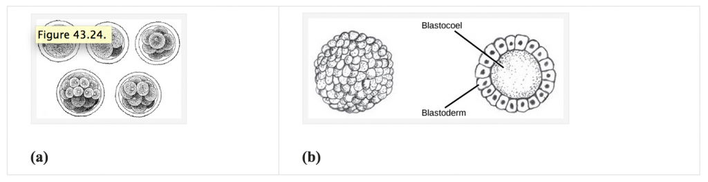
- This stage leads to the formation of a blastula which is a sphere of cells surrounding a fluid-filled cavity called the blastocoel.

Figure 4: Notice how in (a) the zygote undergoes rapid cell divisions to increase the number of cells without increasing its size. Also, look at (b) how the divided cells quickly rearrange themselves to form a hollow ball wherein the cavity is filled with fluid and called blastocoel and the surrounding layer of cells is called blastoderm. Image Credit: Gray’s Anatomy (a) and Pearson Scott Foresman (b) - There are two types of cleavage based on the extent to which cells divide. When the complete division of cells occurs, it is called “holoblastic cleavage” and when the incomplete division of cells occurs, it is called “meroblastic cleavage”. In mammals particularly, holoblastic rotational cleavage occurs. In this type of cleavage,

Figure 5: The cleavage pattern of the early mammalian embryo displays a unique set of divisions. Image Credit: Martin Wuhr - The 1st cleavage: A normal meridional division in the zygote forming 2 blastomeres.
- The 2nd cleavage: One blastomere undergoes meridional division and the other blastomere undergoes equatorial division.
- During the mammalian cleavage stage, the embryo begins to transform. It doesn’t change in its overall size. It is only the cell number that rapidly changes as the division process happens at its full throttle. This leads to an overall reduction in the sizes of the individual cells and an increase in the number of total cells.

Figure 6: Notice how the cleavage happens along cleavage plane I and cleavage plane II in Echinoderms and Amphibians. This is called a radial cleavage pattern. On the other hand, Mammals display rotational cleavage patterns. The first cleavage division in mammalian zygote happens along the cleavage plane I (meridional division), and then after that, one blastomere divides along the cleavage plane IIA (meridional division) and the other blastomeres divide along the cleavage plane IIB (equatorial division). Image Credit: Gilbert SF
-
Blastula Stage
The blastula stage in mammals is a critical milestone in embryonic development as it marks the culmination of intricate processes that transform a fertilized egg into a multicellular structure. Particularly in mammals, the blastula is called “blastocyst”.
The blastula stage represents a remarkable period of preparation for the next phases of development as it sets the stage for the establishment of fundamental tissue layers and eventually leads to organ formation.
Some important pointers to the blastula (here called a blastocyst) stage of embryonic development in mammals are:
- In this stage, the embryo undergoes significant morphological changes and the unique arrangement of cells within the blastula lays the groundwork for the complex journey of growth and differentiation that lies ahead.
- Understanding the characteristics and significance of the blastula stage is crucial for unraveling the intricacies of mammalian development and gaining insights into the mechanisms that shape the building blocks of life.
- The blastocyst stage is a pivotal phase during the early development of mammals as it marks a crucial shift from initial cell divisions to the emergence of distinct cell lineages.
- This stage becomes evident after a series of cell divisions during cleavage which transforms the morula (a solid mass of cells) into a more organized structure.
- At the blastocyst stage, a unique arrangement of cells is observed which is primarily composed of two distinct cell types: the “trophoblast cells” forming the outer layer and the “inner cell mass (ICM)”.
- The trophoblast cells create a protective outer layer that interacts with maternal tissues upon implantation. This interaction facilitates vital processes such as nutrient exchange and waste elimination. Additionally, the trophoblast cells play a vital role in the subsequent development of the placenta, a critical organ that supports fetal growth and development.
- The ICM contains cells with heightened developmental potential. These cells act as precursors to the embryo and eventually give rise to the diverse range of tissues and organs that form the entire organism. The ICM’s pivotal role in generating the three primary embryonic germ layers—the ectoderm, mesoderm, and endoderm—provides the foundation for establishing the fundamental body plan of the developing mammal.
- The distinct cell populations within the blastocyst and their interactions during implantation provide the structural basis for the intricate growth and specialization that characterize the later stages of mammalian development.

-
Gastrula Stage
The gastrula stage in mammalian embryonic development signifies a transformative phase characterized by the “establishment of fundamental germ layers” through a remarkable process known as gastrulation. Following the preceding blastocyst stage, the gastrula stage marks a critical juncture where the developing embryo undergoes intricate morphological and structural changes. By delving into the characteristics and significance of the gastrula stage, one can gain insights into the intricacies of mammalian development and the marvels of how life’s blueprint is intricately woven.
Some important pointers to the blastula (here called a blastocyst) stage of embryonic development in mammals are:
- The gastrula stage is the phase after the blastocyst stage representing a pivotal milestone in the progression of mammalian embryonic development.
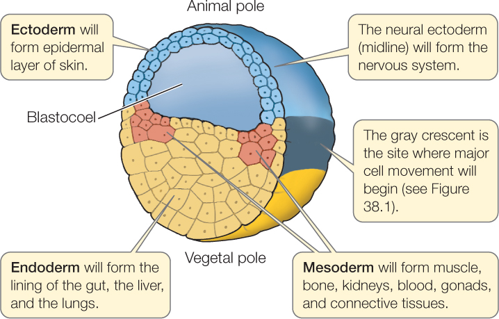
- A central and defining feature of the gastrula stage is the establishment of the 3 primary germ layers: ectoderm, mesoderm, and endoderm. These germ layers are foundational giving rise to diverse tissues and organs within the developing organism.
- Gastrulation is the dynamic process by which these germ layers are initiated. It involves a reorganization of cells within the blastocyst resulting in the spatial arrangement of distinct cell populations and layers.
- The outermost germ layer is known as the ectoderm. It goes on to become the source of critical structures including the skin, nervous system components like the brain and spinal cord, and sensory organs. Occupying the middle position, the mesoderm contributes to vital tissues such as connective tissues, muscles, bones, blood vessels (and blood cells), and the heart. The innermost layer is the endoderm. It shapes the lining of internal organs like the gastrointestinal tract, respiratory system, and liver.
-

Figure 8: The 3 germ layers established in the gastrula stage are vital precursors for the different organs and organ systems. Image Credit: Macmillan Learning - The emergence of the primitive streak is a prominent aspect of gastrulation. This linear structure on the embryo’s surface is the epicenter of cellular migration and the genesis of germ layers.
- Cells embark on a migratory journey from the embryo’s surface to its interior during gastrulation. This orchestrated movement is instrumental in establishing germ layers and sculpting the embryo’s structural basis.
- The gastrula stage fosters interactions among various cell types and tissues which fosters the inception of key developmental cues and patterns.
- The foundational establishment of germ layers during the gastrula stage paves the way for the forthcoming intricacies of tissue and organ patterning and differentiation in the organogenesis stage.
- The gastrula stage is a shared phenomenon across a spectrum of animal species. This underscores its fundamental significance in the broader context of embryonic development.
- From its origins as a relatively uncomplicated blastocyst, the gastrula stage heralds the transition to a more intricate embryonic form characterized by the inception of specialized cellular layers and the potential for diverse tissue generation.
-

Figure 9: Formation of the primitive streak in higher vertebrates during gastrulation. Image Credit: Alexander Minin
-
Organogenesis Stage
The stage of organogenesis in mammalian embryonic development is a transformative period marked by the intricate formation of the organs that will define the future organism. Following the gastrula stage where the foundation of germ layers is laid, organogenesis emerges as a complex process where these germ layers differentiate and give rise to the intricate structures that make up various organs. This phase is a dynamic symphony of cell differentiation, migration, proliferation, and tissue interactions, culminating in the creation of essential organs such as the heart, lungs, brain, and more.
Organogenesis represents the culmination of the embryonic journey, thereby shaping the intricate anatomy and body functions of the developing mammal.
There is a characteristic phylotypic period where the early embryonic stages of vertebrates show higher morphological and molecular resemblance amongst each other. This is reasoned to the higher expression of:
- Evolutionary oldest genes
- Genes under the strongest purifying selection
- Genes with similar temporal expression patterns
There is an interesting paper by Irie, N & Kuratani, S (2011) where they performed a comparative transcriptome analysis at the organogenesis stage for different vertebrates during their phylotypic period. Resemblance at the molecular level was reported. (Ref-9)

Plant Embryonic Development
Inside the seed’s structure, it is the embryo that contains the rudimentary plant. Plant embryos and their embryonic development offer a fascinating glimpse into the intricate process through which a single fertilized cell evolves into a complex multicellular organism.
Unlike animals, plants do not have a defined morula or blastocyst stage, but their embryonic journey is equally captivating. Embryogenesis in plants involves a series of carefully orchestrated events that give rise to the diverse structures and tissues found in adult plants. From the establishment of the embryonic axis to the formation of cotyledons, leaves, stems, and roots, the journey of plant embryos is a testament to nature’s ingenuity in generating life.
A typical dicot plant embryo goes through the globular, heart, torpedo, and final stages.
- Initially, the zygote divides mitotically, forming a proembryo (a mass of undifferentiated cells) anchored to the micropyle by a suspensor.
- As the proembryo divides, it forms a sphere which at this stage is referred to as the globular stage (the first embryo stage or the embryo proper).
- The embryo continues to grow and develop, forming a heart-shaped structure with a prominent embryonic axis and two cotyledon primordial, marking the heart stage.
- Next to this is the torpedo stage where the cotyledons elongate while the base thickens resulting in the embryo assuming a torpedo shape.
- The final stages include events where the embryo further develops its characteristic structures to ultimately develop into a seed (i.e., mature embryo surrounded by protective layers, the seed coat).

Exploring the stages and mechanisms of plant embryonic development provides a deeper understanding of how plants generate and organize their structures, adapt to their environment, and perpetuate their species.
Table 1: A general table of comparison for plant and animal embryonic development. | ||
|---|---|---|
| Aspect | Plant Embryonic Development | Animal Embryonic Development |
| Initiation of Embryo | Fertilization of the haploid ovule by pollen forms a zygote. | Fertilization of the egg by sperm forms a zygote. |
| Germ Layers | No distinct germ layers. | Distinct germ layers (ectoderm, mesoderm, endoderm). |
| Tissue Formation | Differentiation of embryonic tissues from apical cells. | Germ layers give rise to diverse tissues. |
| Embryo Nutrition | Nutrient-rich endosperm supports embryo growth. | Maternal nutrient exchange or yolk sustains the embryo. |
| Embryonic Stages | Progression through globular, heart, and torpedo stages. | Cleavage (morula), blastula, gastrula, organogenesis. |
| Organs and Structures | Cotyledon stems, leaves, and roots develop. | Complex organs and systems develop. |
| Cell Division Pattern | Asymmetric division; apical and basal cells form. | Various patterns of cell division and specialization. |
| Embryo Attachment | Inside archegonium or on the parental gametophyte. | Inside the uterus or eggshell, depending on the species. |
| Placental Connection | Not applicable; no placental connection. | Present in viviparous animals; facilitates nutrient exchange. |
| Embryonic Growth Suspension | Dormancy until germination; reliance on stored nutrients. | Dormancy or continuous growth, depending on species. |
Data Source: Dr. Harpreet Narang of Biology Online
Research And Technology
Embryo research and technology stand at the forefront of scientific exploration with the potential to unveil profound insights into the origins of life and potential avenues for medical advancement. Cutting-edge techniques enable the meticulous study of embryonic development. This helps in unraveling the intricate cellular processes that shape organisms. From groundbreaking discoveries in stem cell therapies to elucidating the genetic underpinnings of complex diseases, embryo research offers a window into the mysteries of existence.
Technological strides such as CRISPR-Cas9 gene editing have empowered scientists to delicately “manipulate embryonic genomes” which look promising for transformative treatments and genetic interventions. This frontier, however, raises ethical questions, hence necessitating a careful balance between innovation and ethical considerations. As we journey into the uncharted territories of embryo research and technology, we navigate not only the realms of science but also the realms of ethics and morality.
-
Biological processes
Studies on embryos from both plants and animals have been ongoing across the globe. Scientists and researchers dealing with embryo research usually seek answers to questions related to developmental biology, evolution and development, cell division, stem cells, and gene expression.
There are several examples of significant scientific discoveries that were made by studying embryos:
- Spemann-Mangold Organizer: Research on amphibian embryos revealed the Spemann-Mangold organizer which is a group of cells orchestrating embryonic development by influencing neural tissue and body axis formation.
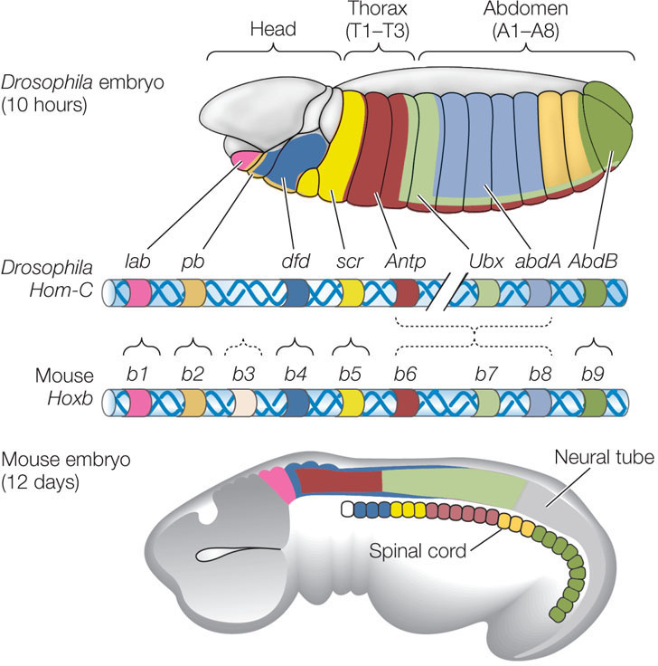
- Hox Genes and Body Segmentation: Drosophila fly embryos research unveiled Hox genes’ role in governing body structure segmentation along the anterior-posterior axis which further shed light on the genetic mechanisms behind segment development.
- Stem Cell Potential: A study of early embryos highlighted pluripotent embryonic stem cells which revolutionized regenerative medicine and therapies reliant on stem cell applications.
- Evolutionary Insights: Comparative embryo studies established evolutionary links and conserved developmental paths which have revealed the shared ancestry and shaped knowledge of species evolution.
- Gene Regulation and Expression: Embryonic research deciphered gene regulation patterns, hence illustrating how genes activate or deactivate during development and eventually guide organ and tissue formation.
- Cell Fate Determination: Embryo investigations unveiled cell fate and differentiation mechanisms which have uncovered the complex processes driving cell specialization.
- Organogenesis and Morphogenesis: Embryo studies illuminated organ and tissue development. This has unveiled intricate processes driving formation, structure, and function.
- Developmental Disorders: Embryo research deepened insights into developmental disorders, thereby identifying genetic and environmental contributors to birth defects.
- Germ Cell Origins: Embryonic study shed light on germ cell origins which are crucial for reproductive biology and assisted reproductive technologies.
- Gastrulation Mechanisms: Embryonic insights elucidated gastrulation which is crucial for tissue differentiation and body structure formation.

-
Assisted reproductive technology
Assisted Reproductive Technology (ART) revolutionizes family planning by aiding couples struggling with fertility challenges. This innovative approach encompasses techniques like in vitro fertilization (IVF), intracytoplasmic sperm injection (ICSI), and gamete donation. ART provides newfound hope to individuals seeking parenthood as it offers solutions when natural conception is elusive. This technology involves controlled ovarian stimulation, egg retrieval, fertilization in a controlled environment, and subsequent embryo transfer. ART has empowered countless families worldwide by bridging the gap between biological limitations and the dream of having a child. Its ethical considerations and continuous advancements underscore its transformative impact on modern reproductive medicine.

-
Cryoconservation of plant and animal biodiversity
Cryoconservation safeguards the rich availability of plant and animal biodiversity by preserving living organisms at “ultra-low temperatures”. This revolutionary technique involves freezing cells, tissues, or even whole organisms which allows their genetic heritage to endure for future generations.
By maintaining biological diversity, cryo conservation plays a pivotal role in conserving endangered species, preserving genetic resources, and facilitating scientific research. This method offers a lifeline to species threatened by habitat loss and climate change. This ensures their survival and potential for future restoration.
Cryo conservation has emerged as a powerful guardian of Earth’s biological legacy by merging science, technology, and conservation in an endeavor to safeguard life’s intricate marvels.

NOTE IT!
“Morula Stage”
The morula stage in mammalian embryonic development signifies a pivotal phase characterized by the compact arrangement of cells following initial rounds of cell division. The word “morula” is derived from the Latin word “morula” meaning ‘mulberry’. This developmental stage showcases a cluster of blastomeres; wherein individual cells formed through cleavage exhibit a distinctive arrangement “resembling the texture of a mulberry fruit”, hence aptly named after it.
The morula stage serves as a vital bridge between the zygote stage and the subsequent blastocyst stage.
During this stage, notable processes like “compaction” and “early signs of cell differentiation” emerge which sets the groundwork for the complex journey of embryonic development.
Understanding the characteristics and significance of the morula stage provides insights into the intricate mechanisms that shape the earliest phases of mammalian life.
Some important pointers to the morula stage of embryonic development in mammals are:
- Time: The time taken for the formation of morula by cleavage process is the longest for the mammalian zygotes as compared to the rest of the animal kingdom; it takes about 12 to 24 hours.
- Initial Division: Following fertilization, the morula stage emerges as a result of the initial rounds of cell division known as cleavage. These divisions give rise to a compact cluster of cells referred to as the morula.
- Cell Cluster: The morula comprises a collection of blastomeres; individual cells formed through cleavage. While the exact count of blastomeres may vary between species, it typically ranges from around 16 to 32 cells.
- Compact Structure Formation: During the morula stage, a process called “compaction” occurs where blastomeres tightly adhere to each other. This compacting results in a more organized and closely united cellular structure.
-

Figure 15: Compaction is a significant and characteristic phenomenon in the early embryonic development of mammals. Time-lapse cinematography (TLC) is a wonderful way to capture and track the progress of such biological phenomena with precision in real-time. These timed shots show how the human embryo divides rapidly to yield several blastomeres (a-e), followed by the flattening of blastomeres (f) and intercellular boundaries becoming obscure (g-i). It finally reaches a stage where all blastomeres unify in one cluster (j-k). Image Credit: Kyoko Iwata - Potential of Blastomeres: Blastomeres in the morula stage possess “totipotency” implying that each cell can develop into an entire organism if isolated.
- The precursor to Blastocyst: Changes and differentiation take place within the morula to give rise to the blastocyst.
- Timing and Transience: The morula stage occurs several days after fertilization and is a transient phase within embryonic development.
- Developmental Significance: The morula stage is pivotal as the embryo progresses from a solitary zygote to a multicellular structure with distinct cells that will contribute to various tissues and organs.
- Cell Specialization Onset: Even though full differentiation hasn’t occurred, some blastomeres in the morula stage begin to display early signs of specialization.
- Consistency Across Species: The morula stage is a shared feature among various mammalian species, underscoring its significance in the initial stages of development.
- Stem Cell Exploration: Researchers have taken an interest in stem cells derived from the morula due to their potential to differentiate into diverse cell types. This holds promise for “applications in regenerative medicine”.
- Ethical Dimensions: The morula stage raises ethical considerations concerning embryonic research, assisted reproductive technologies, and the commencement of human life.
-

Figure 16: Stages of Stem Cell Potential: From totipotent morula cells with total developmental versatility, to pluripotent inner mass cells forming body tissues, multipotent stem cells specializing closely, and self-renewing unipotent cells. These are promising avenues for regenerative medicine but with some ethical conundrums that need to be dealt with care. Image Credit: Molecular Diagnostic Service
Take the Embryo – Biology Quiz!

Further Reading
References
- Gilbert SF. Developmental Biology. 6th edition. Sunderland (MA): Sinauer Associates; 2000. Early Mammalian Development. Available from: https://www.ncbi.nlm.nih.gov/books/NBK10052/
- Yamanaka, Y., Ralston, A., Stephenson, R. O., & Rossant, J. (2006). Cell and molecular regulation of the mouse blastocyst. Developmental dynamics: an official publication of the American Association of Anatomists, 235(9), 2301-2314.
- Hasley, A., Chavez, S., Danilchik, M., Wühr, M., & Pelegri, F. (2017). Vertebrate embryonic cleavage pattern determination. Vertebrate Development: Maternal to Zygotic Control, 117-171.
- Iwata, K., Yumoto, K., Sugishima, M., Mizoguchi, C., Kai, Y., Iba, Y., & Mio, Y. (2014). Analysis of compaction initiation in human embryos by using time-lapse cinematography. Journal of assisted reproduction and genetics, 31, 421-426.
- Saini, A. K., Saini, R., Bansode, H., Singh, A., & Singh, L. (2020). Stem Cells: A Review Encompassing the Literature with a Special Focus on the Side-Lined Miraculous Panacea; Pre-Morula Stem Cells. Current stem cell research & therapy, 15(4), 379–387. https://doi.org/10.2174/1574888X15666200311141731
- Strelchenko, N., Verlinsky, O., Kukharenko, V., & Verlinsky, Y. (2004). Morula-derived human embryonic stem cells. Reproductive biomedicine online, 9(6), 623–629. https://doi.org/10.1016/s1472-6483(10)61772-5
- Zakrzewski, W., Dobrzyński, M., Szymonowicz, M. et al. Stem cells: past, present, and future. Stem Cell Res Ther 10, 68 (2019). https://doi.org/10.1186/s13287-019-1165-5
- Chernoivanenko, I. S., Minin, A. A., & Minin, A. A. (2013). Role of vimentin in cell migration. Russian Journal of Developmental Biology, 44, 144-157.
- Irie, N., & Kuratani, S. (2011). Comparative transcriptome analysis reveals vertebrate phylotypic period during organogenesis. Nature communications, 2, 248. https://doi.org/10.1038/ncomms1248
- dos Santos-Neto, P. C., Cuadro, F., Souza-Neves, M., Crispo, M., & Menchaca, A. (2023). Refinements in embryo manipulation applied to CRISPR technology in livestock. Theriogenology.
- Graham, M. E., Jelin, A., Hoon Jr, A. H., Wilms Floet, A. M., Levey, E., & Graham, E. M. (2023). Assisted reproductive technology: Short‐and long‐term outcomes. Developmental Medicine & Child Neurology, 65(1), 38-49.
- Kaviani, B., & Kulus, D. (2022). Cryopreservation of Endangered Ornamental Plants and Fruit Crops from Tropical and Subtropical Regions. Biology, 11(6), 847. https://doi.org/10.3390/biology11060847
©BiologyOnline.com. Content provided and moderated by Biology Online Editors.


:max_bytes(150000):strip_icc()/2795073-stages-of-prenatal-development-01-5a3040f6eb4d5200362d5553.png)