
Anaphase
n., plural: anaphases
[ˈænəˌfeɪz]
Definition: The separation of chromosomes, i.e., the separation of chromatids in mitosis and in meiosis II or the separation of homologous chromosomes in meiosis I
Table of Contents
In order to keep the process of biological existence ever-going, the phenomenon of cell multiplication is very important. Cells don’t just multiply in one go, they do so via defined steps and phases. The biological world is a complex amalgam of several cellular and molecular level steps. Innumerable small steps acting in a synchronized manner pave way for a cell to produce more of its kind. This, in totality, ensures the continuation of life forms on the face of this planet. When we talk about eukaryotic life forms, there are two biological processes acting at cellular levels; they are mitosis and meiosis. Both of these processes give rise to new cells from the parent cell. Let’s delve deeper into the topic and learn to answer questions, such as: what is anaphase? … what are sister chromatids? … how do the chromosomes separate in anaphase I? … are sister chromatids identical? … how many cells are there in anaphase?
Anaphase Definition
In biology, anaphase is the stage of cell division where the chromosomes from the metaphase split apart leading to their movement to the opposite poles of the cell. Anaphase, etymologically, is derived from two Greek words, where ‘ana’ means ‘backward‘ and ‘phasis’ means ‘appearance‘.
The two copies of chromosomes are formed during the DNA replication of the S phase of the cell cycle. When the cell cycle progresses to cell division, the duplicated chromosomes go through the various stages in the following order: prophase, metaphase, anaphase, and telophase. Anaphase occurs after metaphase and before telophase. Metaphase is the stage wherein the chromosomes are found at the metaphase plate. The separation of chromosomes to sister chromatids happens next. Once separated, each chromatid is now referred to as a daughter chromosome. This separation process marks the significantly important anaphase and is its characteristic feature of it and so that by telophase the spindle disappears and two nuclei form.

Fundamental concepts
- Mitosis is the process by which all somatic cells produce two daughter cells that are genetically identical in nature. Mitosis cell division occurs in all unicellular and multi-cellular eukaryotic cells. Mitotic cell division doesn’t happen in prokaryotic cells.
- Meiosis is the process by which germinal or germ cells produce four daughter cells that are genetically dissimilar and in which the chromosomes are reduced by half. This leads to the formation of meiospores or sexual spores. Meiosis occurs only in the sexually-reproducing eukaryotes.
Essentially, both mitosis and meiosis consist of an ordered chain of steps or phases namely prophase (first step), metaphase, anaphase, and telophase (last step). This article will focus mainly on the discussion of anaphase.
An important point to note here is that there’s only one anaphase in the mitosis process while there are two anaphases in the meiosis process (anaphase I and anaphase II).
Important pointers
Some important pointers about anaphase are:
- This phase happens only once in mitosis while it happens twice in the meiosis process.
- This phase is characterized by microtubules’ disintegration.
- This phase is when the chromosomes reach their overall maximum condensation.
- This phase is marked by chromosome segregation (the sister chromatids are moving apart) and the subsequent re-formation of the nuclear membranes and the nucleoli.
- This phase is characterized by the disassembly of tubulin fibers leading to their shortening and eventually pulling apart of the sister chromatids to the opposite poles.
- This phase is distinctively noted by the appearance of V-shaped structures. So, when asked, ‘what does anaphase look like’, we can provide a decent explanation. These V-shaped structures are none other than the sister chromatids which are being pulled apart from the metaphasic plate. When you are trying to identify the cell division phase, specifically, anaphase under the microscope, you should be looking out for V-shaped structures.

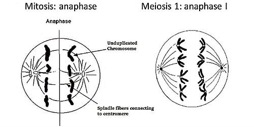
Figure 2: Anaphase image. Source: Delta Vision Roy van Heesbeen, WikiMedia. - In the case of mitosis (anaphase), “sister chromatids” separate.
- In the case of meiosis (anaphase I), “homologous chromosomes” separate.
- In the case of meiosis (anaphase II), “sister chromatids” separate.
- So, it must be clear now that sister chromatids separate during mitosis anaphase and meiosis anaphase II.


Figure 3: Please notice that sister chromatids are pulled apart in anaphase of mitosis while homologous chromosomes are separated in anaphase I of meiosis. The diagram also shows the star-like structures. They are the astral microtubules that radiate towards the cell membrane from the centrosomes. Centrosomes are structures containing a pair of barrel-shaped centrioles and are found only in animal cells, not in plant cells. Other microtubules that radiate from the centrosomes are interpolar (or simply polar) microtubules (radiating towards the mitotic spindle and in between poles) and kinetochore microtubules (connect directly to kinetochores). Image Credits: QS Study (left) and E.V. Wong, Bio.Libretexts.org. - The ploidy level at the mitosis anaphase is 4n.
- The ploidy level at the meiotic anaphase I is 2n.
- The ploidy level at the meiotic anaphase II is n.
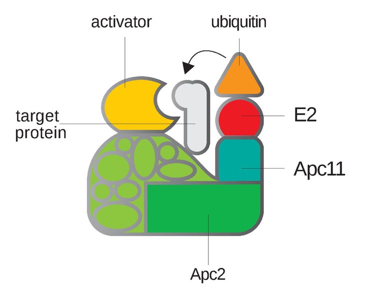
- Role of APC: Early anaphase is marked by ubiquitination of the “securin” protein by the APC (or anaphase-promoting complex). The securin protein is inhibitory in nature. It inhibits one enzyme known as separase. Separase enzyme (a type of protease) performs an essential role in breaking down another protein known as cohesin. Cohesin is actually responsible for keeping the sister chromatids held together. As the APC gets active, it ubiquitinates the securin. As securin is removed from the picture, the control over the separase enzyme is uplifted. As separase gets the freedom to act, it acts immediately upon cohesion protein. The activity of separase breaks down cohesion and the sister chromatids can’t be held together anymore. Finally, the sister chromatids are separated by this complex interplay of events.

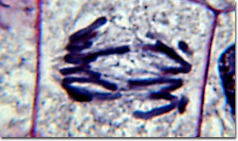
Figure 4: Early anaphase can be noticed in this photomicrograph. We can clearly and distinctly see the microtubules and the separating homologous chromosomes. Image Credit: The Florida State University Figure 5: Late anaphase can be noticed in this photomicrograph. We can clearly see the cell proceeding towards telophase. Image Credit: Florida State University
Watch this vid about anaphase:
Anaphase is the phase following metaphase and preceding telophase of cell divisions (in both mitosis and meiosis). It is highlighted by the separation and movement of chromosomes from the metaphase plate towards the poles of the spindle.
Quick Overview: Cell divisions in eukaryotes, particularly mitosis and meiosis, are important since they give rise to new cells. Mitosis produces two cells that are genetically identical. Meiosis produces four cells that are genetically dissimilar and in which the chromosomes are reduced by half. Both mitosis and meiosis are comprised of chronological phases: (1) prophase, (2) metaphase, (3) anaphase, and (4) telophase. Since meiosis is comprised of first and second meiotic divisions, these phases occur twice, each designated as I and II.
Mitotic anaphase: After the mitotic metaphase (when all the chromosomes align at the metaphase plate and microtubules attach to the kinetochores), the succeeding phase is anaphase wherein the chromosomes would now move towards the poles of the spindle.
Meiotic anaphase: In meiosis, anaphase occurs twice, i.e. anaphase I at the first meiotic division and anaphase II at the second meiotic division.
Etymology: from the Latin or Greek aná, meaning “back” and phase, phásis, meaning “appearance”.
Anaphase in Mitosis
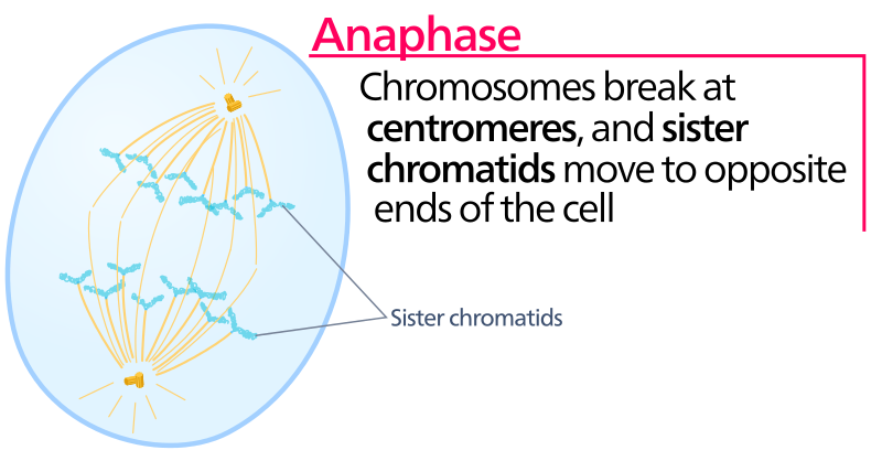
Anaphase of cell mitosis is the third phase of the active cell division process. It happens after the prophase and metaphase stages. Sister chromatids are joined at the centromeres; they need to be separated next. After the spindle fibers are already arranged and properly attached to the chromosomes at their centromeres which are lined up neatly at the equatorial plane, anaphase begins. The coordinated disintegration of the microtubules follows and the tubulin fibers start disassembling.
So, what happens during anaphase? It is when:
- The chromatids that make up each chromosome are strategically pulled apart in order to ensure the daughter cells that are bound to form later, receive their individual set of separate chromosomes.
- The chromatids from the metaphasic plate after separation in the anaphase stage are now considered individual chromosomes.
- After they reach their opposite poles, anaphase progresses, and the telophase begins.
- You must be wondering what moves the chromatids during mitosis, so it’s the mitotic spindle fibers.

Anaphase in Meiosis
Anaphase in meiosis occurs twice; anaphase I and anaphase II. The characteristic features of both anaphase stages are different. At the meiotic metaphase I equiplate, homologous chromosomes are lined up, so it’s essentially the homologous chromosomes that separate in the anaphase I stage and not the sister chromatids as expected. Let’s look at each one of them individually.
Anaphase I
Definition of anaphase 1: the first anaphase out of the two anaphases of the meiotic division. Even though there is only one round of replication in meiosis (just like mitosis) but there are two rounds of segregation in meiosis. Anaphase I is the first round of segregation. This is the reductional segregation step. As the chromosome number is halved after this segregation, we see the change in ploidy in the final product (gametes). The separation of homologous chromosomes is a landmark event in anaphase I.
Major events of anaphase 1:
- Shortening of kinetochore microtubules
- Homologous chromosomes moving towards opposite spindle poles
- Lengthening of non-kinetochore microtubules
- Centrosomes are pushed far apart
- Elongation of the cell (as a preparation step for division)
- Degradation of cohesin but only from chromosome arms
- No cohesin degradation from around centromere. Reason for the preservation of the cohesin around the centromere: It happens due to the presence of a protein called Shugoshin. Shugoshin shields the cohesin around the centromere and prevents the separation of sister chromatids.
Anaphase II
Definition of anaphase 2: the second anaphase of the two anaphases of the meiotic cell division is anaphase II. Anaphase II is the second round of segregation. This is the equational segregation step. As the DNA content gets halved, this is almost similar to mitosis. The separation of the two sister chromatids is the landmark event in anaphase II.
Major events of anaphase 2:
In this stage, the remaining cohesin is also degraded from the centromere region. The Shugoshin no more protects the cohesin holding the sister chromatids together at the centromere now. This paves way for the sister chromatids’ separation. After their separation, they are called ‘sister chromosomes’ on reaching their respective destined opposite poles.
In animal cells, the events that occur in cell divisions, especially metaphase and anaphase, are driven by motor proteins. They “carry” the chromosomes or other microtubules as they walk along the surface of the microtubules. Their role in animal cell division is crucial. They are responsible for the segregation of chromosomes and they make sure that they do so with the highest fidelity as much as possible.
 |  |
| Various motor proteins “walk” and carry cargo at the microtubules, indicating their modulation of microtubular kinetics. Motor proteins, particularly dyneins and some kinesins, walk toward the minus ends of microtubules anchored at the spindle pole, which could account for the chromosomal movement during anaphase. Image Credits: Wordeman, L. (2010). How kinesin motor proteins drive mitotic spindle function: Lessons from molecular assays. Seminars in Cell & Developmental Biology, 21(3), 260–268. (left) and E.V. Wong, Bio.Libretexts.org (right). | |
Watch this vid of a molecular structure called kinesin, a motor protein walking on the surface of a microtubule while carrying a ‘cargo’.
Relation to the cell cycle
In the cell cycle, the longest phase is the interphase composed of G1, S, and G2 phases. All these together capture almost 95% of the cell cycle’s time. Only 5% time is allotted to M-phase. M-phase consists of prophase, metaphase, anaphase, and telophase. If looked closely, anaphase is left with only a little 1% of the cell cycle’s duration.
The role of APC is essential at the beginning of the anaphase. This intricately regulated transition from metaphase to anaphase has many players acting at the molecular level.
 Many genes share some essential roles and functions in the cell cycle progression and division. Even in anaphase, there is a long list of genes involved like APC, BUB1B, CDC14A, CDH1, FZR1, MAD2L1, MXI1, PROC, RAD21, etc. The functioning of anaphase-promoting complex (APC) as explained above is irreplaceable. For a cell to transition from metaphase to anaphase, the gene of APC has to function properly in order to make a functional mRNA and an APC protein. APC is essential for the transition from metaphase to anaphase. Source: The Cell Cycle. Principles of Control.
Answer the quiz below to check what you have learned so far about anaphase.

Further Reading
References
- Castro, A., Bernis, C., Vigneron, S. et al. The anaphase-promoting complex: a key factor in the regulation of cell cycle. Oncogene 24, 314–325 (2005). https://doi.org/10.1038/sj.onc.1207973
- Peters, JM. The anaphase promoting complex/cyclosome: a machine designed to destroy. Nat Rev Mol Cell Biol 7, 644–656 (2006). https://doi.org/10.1038/nrm1988
- The Cell Cycle. Kimball’s Biology Pages.
- Schafer KA (November 1998). “The cell cycle: a review”. Veterinary Pathology. 35 (6): 461–78. doi:10.1177/030098589803500601
©BiologyOnline.com. Content provided and moderated by Biology Online Editors.

