
Telophase
n., plural: telophases
[ˈtɛləˌfeɪz]
Definition: The final stage of mitosis or meiosis
Table of Contents
Telophase is the stage of cell division characterized by the decondensation of chromosomes, and the nuclear envelope assembly around each set of chromosomes. After telophase, the cytokinesis process occurs resulting in the formation of 2-4 daughter cells depending on cell division type (mitosis/meiosis). This marks the completion of chromosome segregation and the beginning of interphase or the next round of division.
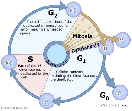
The cell cycle is a fascinating process that has mesmerized scientists of all domains, particularly cell biology scientists. Let’s give you a brief overview before we dive into the ‘intricacies of telophase’; a short phase in the lengthy dividing cell timeline, i.e., the cell cycle process!
When we unlock the secrets of how our bodies grow, heal and function at the microscopic level, we come face to face with the smallest entities of life called cells. Just imagine your body to be a bustling city with millions of citizens called ‘cells’; each one of them with their own job and responsibilities. Now each of these citizens also has their own life journey. This is analogous to each of the cell’s life journeys that we scientifically call the ‘cell cycle’.
As all citizens go about in their well-choreographed life routine, so do all the cells by the medium of the cell cycle!
Each phase of the cell cycle is as important as the routine or scheduled job of each citizen at any given hour of the day. This brings us to the most important point of today’s discussion “not even a single phase of the cell cycle can be replaced or omitted”. Of all the different yet important phases of the cell cycle, one is “TELOPHASE”! This lesson will talk in depth about telophase, its presence in both mitosis and meiosis types of cell division, and its peculiarities. So, read on to gain deeper insights into this phase of the cell cycle.

Telophase Definition
Telophase can be defined as the phase common to both mitosis and meiosis that’s characterized by:
-
- For Mitotic Cell: Decondensation of chromosomes as chromosomes begin to transform into chromatin, breakage of now-defunct spindle fibers apparatus, formation of new nuclear envelopes for nuclear material of two daughter cells, and final preparation for cytokinesis.
- For Meiotic Cell: Telophase I and Telophase II of meiosis have different characteristic features.
-
Timing of mitotic and meiotic telophases
- The mitotic telophase is preceded by anaphase and followed by cytokinesis (where the cytoplasm physically divides to form two daughter cells).
- The meiotic telophase I is preceded by anaphase I followed by short cytokinesis and prophase II. And the meiotic telophase II occurs during the second meiotic division and is preceded by anaphase II and followed by cytokinesis.

-
Telophase Etymology
The term telophase is derived from 2 Ancient Greek words;
- The first word “τέλος” that is “télos” meaning ‘the end, the completion, or the result’.
- The second word “φάσις” that is “phásis” meaning ‘the appearance’.
NOTE IT!
Importance of Telophase in the Cell Division and Cell Cycle Process
Since in the life of any somatic cell, the telophase is the last phase of the M-phase (mitotic phase), completes a full circle for the eukaryotic cell (plant cells or animal cells). Once the cell leaves telophase, it is ready for the division of its cytoplasmic content through the process of “cytokinesis” leading to the creation of two daughter cells. This highlights the underlying importance of telophase in the life of a somatic cell.

-
For the reformation of functional cytoplasmic structures
When we observe the basic characteristics of telophase, we also observe that telophase “basically reverses all the processes” that were manifested in the preceding prophase and prometaphase. This means that whatever disintegration of nucleolus material and inner nuclear membrane took place in prophase and prometaphase is taken care of in the telophase. New nuclear membranes/envelopes form around the separated chromatin material of the two daughter cells. These envelopes are contributed by the vesicles of the endoplasmic reticulum. Also, new nucleoli are formed for both the daughter cells in the telophase. This step is common for both telophases — telophase of the cell undergoing mitosis and telophase II of the cell undergoing meiosis.

-
Ensures chromosomal segregation and decondensation
Another important point to note is that telophase ensures that the chromosomes (the condensed form of chromatins) are pulled apart to the cell poles and decondensed back to their expanded form, i.e., the chromatin. This step is crucial as the two daughter cells are about to divide in the subsequent cytokinesis phase and form two separate functional identities. For genomic content to stay viable, it must be converted to the chromatin state.
-
Restoration of cellular organization
Telophase is also marked by the disassembly of the mitotic spindle apparatus. As the chromosomes change their state to a more expanded form of chromatin, it’s important that no spindle microtubules unnecessarily lie within the cell. This is why telophase is accompanied by depolymerization of these spindle microtubules.
Though the length of the telophase is merely 2% of the total cell cycle’s duration, its importance in cell division and cell cycle should not be ignored. Only after successful telophase, the cell is signaled to leave the M-phase and begin the segregation of cell constituents into the two separate daughter cells.
-
- Does telophase have checkpoints?
In contrast to the earlier phases of the M-phase, the telophase does not have any specific checkpoint/s. We all know that checkpoints are the important regulatory mechanisms of the cell cycle that play an instrumental role in ensuring the proper progress of the cell cycle and the prevention of any errors or abnormalities.
While the lack of checkpoints can be interpreted otherwise, telophase is tightly regulated by the checkpoints in the previous phases of G1, S, and G2 phases. During the telophase, the genetic material has already been segregated into two distinct nuclei and the cell is in the process of physically dividing into two daughter cells through cytokinesis indicating that the cell has completed all the essential steps of mitosis thus explaining the absence of any specific checkpoints in the telophase.
-
Key driver of telophase
The process of “Cdk substrate dephosphorylation” is the key driver of telophase. Cdk stands for cyclin-dependent kinase. As we all know that telophase is the final stage of mitosis and is marked by a critical step of the transformation of separated genetic material into two distinct nuclei. We all notice that the cell cycle is tightly regulated for proper nuclear and cell division by the Cdks and their regulatory proteins, i.e., cyclins. It is this mitotic Cdk-cyclin complex that specifically governs mitosis progression. As telophase marks the end of mitosis, these regulatory networks need to be shut down as their purposes are fulfilled. Sometimes, during the end of the metaphase itself, the activity of mitotic Cdk-cyclin complexes can be noticed to be diminishing. This peculiar decline in the Cdk activity is attributed to the dephosphorylation of Cdk substrates in the late metaphase or entire telophase. If asked to define the dephosphorylation process, we can elaborate it as “the process of removal of phosphate groups from proteins that were serving as key regulatory mechanisms for protein function”.
Importance of dephosphorylation of Cdk substrates:
» It leads to the mitotic spindle disassembly.
» It allows the chromosomes to decondense and reorganizes into two distinct sets.
» It incited the development of new nuclear envelopes.
» It finally ensures the establishment of the 2 separate nuclei of the daughter cells.
Watch this vid about telophase:
Biology definition:
Telophase is the stage of cell division (i.e. mitosis and meiosis) that occurs after the anaphase. It is characterized by the decondensation of chromosomes, and the nuclear envelope assembly around each set of chromosomes. Telophase, together with cytokinesis (the process of cytoplasmic division), marks the final stage of cell division.
Quick Overview:
Cell divisions in eukaryotes — particularly mitosis and meiosis — are important since they give rise to new cells. Mitosis produces two cells that are genetically identical. Meiosis produces four cells that are genetically dissimilar and in which the chromosomes are reduced by half. Both mitosis and meiosis are comprised of these chronological phases: (1) prophase, (2) metaphase, (3) anaphase, and (4) telophase. Since meiosis is comprised of first and second meiotic divisions, these phases occur twice, each designated as I and II.
Etymology: Ancient Greek “τέλος” (télos), meaning “completion”, Ancient Greek “φάσις” (phásis), meaning “an appearance”.
Telophase In Mitosis
Understanding the intricacies of mitotic telophase can help us to garner a greater appreciation for the precision with which the complex cell cycle operates. It allows us to recognize the remarkable mechanisms that cells employ for the growth, development, and maintenance of living organisms. Telophase is of utmost importance in mitosis because it marks the final steps of cell division.
The successful completion of telophase ensures that the genetic material is accurately distributed which allows each daughter cell to receive a complete set of chromosomes. Additionally, the reformation of the nuclear envelope, the formation of nucleoli, and the disassembly of the mitotic spindle contribute to the proper functioning and stability of the daughter cells.

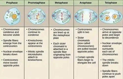
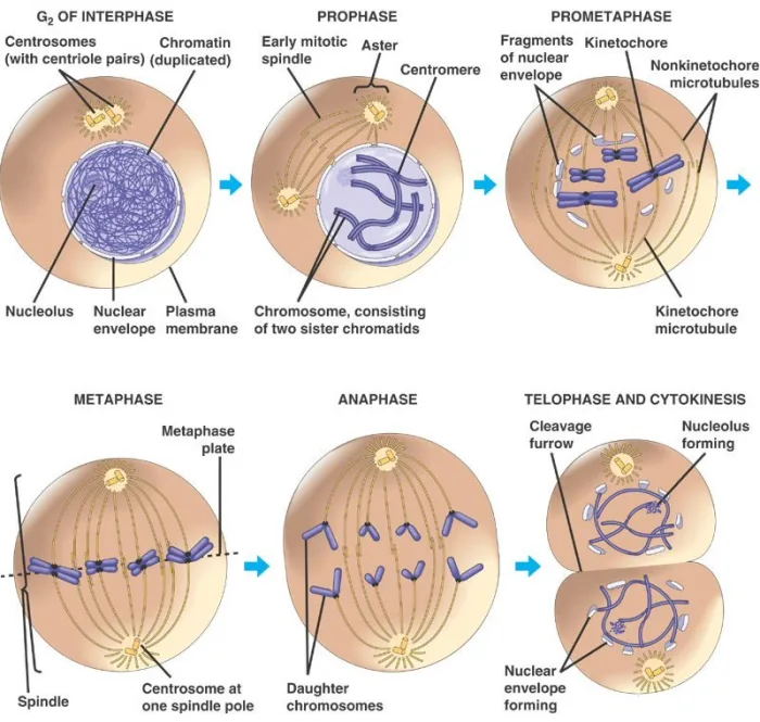
Telophase, being the final stage of mitosis, is marked by:
- Chromosome Decondensation: In earlier stages of mitosis (prophase, metaphase, and anaphase), chromosomes are highly condensed. This allows us to easily visualize them under a microscope in all those stages. However, during telophase, they begin to decondense, allowing the genetic material to relax. This relaxed and expanded state of chromatin is more accessible for gene expression and various cellular activities.
- Nuclear Envelope Reassembly: At the beginning of mitosis (prophase and prometaphase), the nuclear envelope breaks down. But in telophase, this process is entirely reversed, and the nuclear envelope reforms around the separated chromosomes. This reformation is essential for two reasons:
- First: To enclose the genetic material within each daughter cell and protect it from potential damage.
- Second: To re-establish the distinct nuclear compartments and enable specific cellular processes to occur within the nucleus.
Nuclear envelope reformation is accompanied by the binding of the nuclear pore scaffold protein to chromatin. And this is one of the earliest notable events in this reformation process.
- Nucleolus Formation: The nucleolus is a structure found within the nucleus. It plays a crucial role in the production of ribosomes (cells produced protein synthesis bodies). In telophase, new nucleoli start to reappear within each new nucleus. This is significant because it ensures that protein synthesis can resume efficiently in the daughter cells which will allow them to carry out their functions using their proteins.
- Mitotic Spindle Disassembly: A structure called the mitotic spindle helps in the separation of the duplicated chromosomes during the early stages of mitosis. However, in telophase, the mitotic spindle begins to disassemble. This disassembly is important because it allows the chromosomes to relax and spread out within the newly forming daughter cells. It also frees up cellular resources that were previously dedicated to the spindle aiding their redirection for other cellular processes.
- Cytokinesis: Although cytokinesis isn’t strictly a part of telophase, it often overlaps with this stage. Cytokinesis is the physical division of the cell constituents into two daughter cells. It is accompanied by the formation of a contractile ring (composed of actin and myosin) in the middle of the cell. This contraction of the contractile ring causes the cell membrane to pinch (inwards) and leads to the formation of a cleavage furrow. Eventually, this furrow deepens and leads to the wholesome separation of the cytoplasm. This subsequently leads to the formation of two distinct daughter cells with their cell membrane.

Telophase In Meiosis
Telophase in meiosis and mitosis differ in terms of chromosome number, genetic variation, purpose, cytokinesis, and the behavior of centromeres. These differences reflect the distinct goals and outcomes of meiosis (formation of gametes and genetic diversity) versus mitosis (growth, repair, and asexual reproduction).
Telophase in meiosis differs from telophase in mitosis in several key aspects:
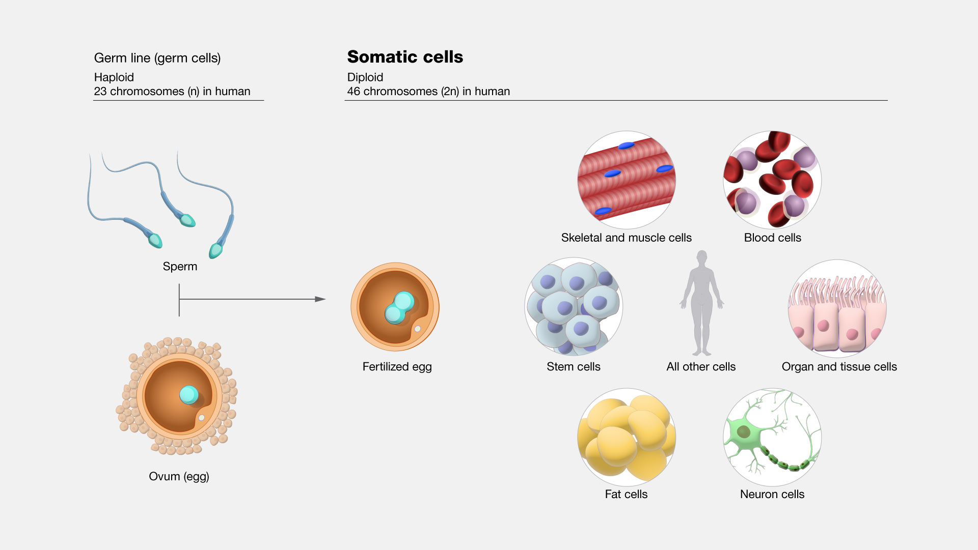
- Chromosome Number: In meiosis, two telophases occur; telophase I and telophase II. Telophase II occurs after ‘two rounds of chromosome segregation’ (meiosis I and meiosis II) which results in the production of haploid cells or gametes. In contrast, in mitosis, telophase occurs after ‘1 round of chromosome segregation’ which results in the production of diploid cells that contain the same number of chromosomes as the parent cell. Note: Somatic cells undergo mitosis whereas germinal cells undergo both mitosis and meiosis.
- Genetic Variation: Telophase in meiosis is critical as it leads to the active generation of genetic diversity. Through processes like “crossing over” and “independent assortment”, homologous chromosomes can exchange genetic material which leads to the shuffling of their genes during meiosis I. This shuffling results in unique combinations of alleles in the chromosomes of resulting daughter cells. Contrary to this, mitosis is not accompanied by any such exchange of genetic material between homologous chromosomes which is why we see no genetically non-identical daughter cells; the daughter cells are exact replicas of parent cells after mitosis!
- Purpose: The purpose of telophase in meiosis is to produce gametes (sperm and eggs) with half the chromosome number of the parent cell. These gametes are necessary for sexual reproduction as they combine during fertilization to restore the diploid chromosome number in the offspring/s. In contrast to that, telophase in mitosis is vital for the completion of cell division to promote the growth, development, and maintenance of multicellular organisms. This happens through the production of genetically identical daughter cells for tissue repair, growth, or asexual reproduction via the process of mitosis.
- Cytokinesis: In meiosis, cytokinesis occurs twice; after both telophase I and telophase II, thereby resulting in the division of the original cell into four haploid cells. On the contrary, in mitosis, cytokinesis occurs only once and typically overlaps with telophase, and involves the division of the original cell into two identical diploid daughter cells.
- Centromere Connection: During telophase in meiosis I, the centromeres of sister chromatids remain connected, while the homologous chromosomes separate. This connection allows the exchange of genetic material between homologous chromosomes. In contrast, during telophase in meiosis II and mitosis, the centromeres of the sister chromatids are no longer connected and this eventually leads to the separation of the sister chromatids.

-
Telophase I
Telophase I and Telophase II in meiosis are distinct stages with specific characteristics and differences. Telophase I is associated with:
- The separation of homologous chromosomes.
- The formation of two diploid daughter cells.
- Carries the potential for genetic variation.
- Occurs after the completion of the first round of chromosome segregation during anaphase I in meiosis I.
- Contrary to common misconception, there is the reformation of the nuclear envelope even after telophase I. This reformation of the nuclear envelope helps in protecting the genetic material and establishment of distinct nuclei within each daughter cell.
- Also, cytokinesis follows telophase I in most organisms. This physical division is often unequal; meaning it results in the formation of 1 cell containing more cytoplasm than the other. And this uneven or non-uniform distribution is necessary for the subsequent stages of meiosis.
-
Telophase II
Telophase II is associated with:
- The separation of sister chromatids.
- The formation of four haploid daughter cells/gametes.
- Occurs after the completion of the second round of chromosome segregation during anaphase II in meiosis II.
- Following telophase II, cytokinesis occurs again. This leads to the division of the two cells from each of the previous divisions into a total of four haploid cells also called the “gametes” or “reproductive cells” that possess half the number of chromosomes found in the original parent cell.
NOTE IT!
Plant Cell Telophase and Cytokinesis
In plant cells, telophase and cytokinesis occur quite differently from animal cells. Although telophase in plant cells is similarly marked by the movement of chromosomes toward opposite poles of the cell with the help of spindle fibers, decondensation of chromosomes, nuclear envelope reformation, and cellular organization restoration, it differs from animal cells in the following:
- Having no centrosomes, plant cells have microtubule-organizing centers that act as an alternative to centrosomes
- Plant cells form phragmoplasts in late metaphase to act as a scaffold for guiding the vesicles carrying cell wall materials to the equatorial region for cell plate formation.
- Having mentioned about cell plate, the plant cell divides not by forming a cleavage furrow (via contractile rings) but by forming a cell plate. The cell plate forms from the fusion of Golgi vesicles containing cellulose and other polysaccharides. As pointed out earlier, these vesicles have moved to the equatorial region of the plant cell as guided by the phragmoplast scaffold. Gradually, the cell plate expands toward the cell edges. One might wonder though where the vesicles have come from if there is no fully-formed Golgi apparatus yet at this point. Remember that the Golgi apparatus have only fragmented and is dispersed in the cytoplasm. These Golgi vesicles would therefore be derived from the fragmented Golgi containing the necessary cell plate components, which during telophase and cytokinesis will assemble at the dividing plane (equatorial region), expand, and eventually fuse with the existing cell wall. The (fragmented) endoplasmic reticulum also has a role in cell plate formation. It supplies membrane components (e.g., phospholipids, proteins, etc.) necessary for the expansion and fusion of Golgi vesicles during cell plate formation.
- While the full restoration of functional organelles, including the Golgi apparatus, can be expected after cell division, particularly during interphase, the reformation of the nuclear envelope in plant cells occurs while the cell plate forms. Similar to animal cells, though, the membrane materials for the nuclear envelope are provided by the ER.
- Thus, in addition to the cell membrane, cell walls are formed during the final stage of cell division of a plant cell.

Take the Telophase – Biology Quiz!

References
- Bower, J.J., Vance, L.D., Psioda, M. et al. Patterns of cell cycle checkpoint deregulation associated with intrinsic molecular subtypes of human breast cancer cells. npj Breast Cancer 3, 9 (2017). https://doi.org/10.1038/s41523-017-0009-7
- Johnson, D. G., & Walker, C. L. (1999). Cyclins and cell cycle checkpoints. Annual review of pharmacology and toxicology, 39.
- Schafer, K. A. (1998). The cell cycle: a review. Veterinary pathology, 35(6), 461-478.
- Israels, E. D., & Israels, L. G. (2000). The cell cycle. The oncologist, 5(6), 510-513.
- Knoblich, J. A. (2010). Asymmetric cell division: recent developments and their implications for tumour biology. Nature reviews Molecular cell biology, 11(12), 849-860.
- Debatisse, M., & Rosselli, F. (2019). A journey with common fragile sites: From S phase to telophase. Genes, Chromosomes and Cancer, 58(5), 305-316.
©BiologyOnline.com. Content provided and moderated by Biology Online Editors.


