Prophase
n., plural: prophases
[ˈprəʊˌfeɪz]
Definition: the first stage of the M-phase of the cell cycle
Table of Contents
Prophase is the first stage of mitosis; the very first step in this crucial process of the M-phase of the cell cycle. Now as we all know that cell divisions are also of two types namely mitosis and meiosis, we shall learn in the following sections about prophase – mitosis, prophase 1 – meiosis, and prophase II – meiosis. Let’s move ahead and try to find answers to some interesting questions, e.g., what is prophase, what happens in prophase 2, what happens during interphase, what happens in mitosis, and so on. The scope of this article is majorly limited to prophase but you should be in a position to answer all these questions by the end of this literary piece.
Prophase Definition
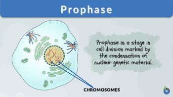
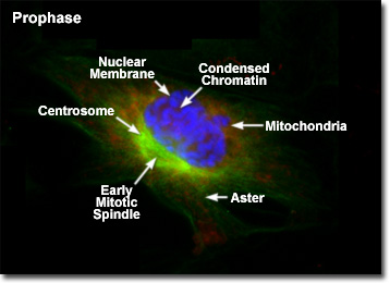
In Biology, the definition of prophase is this: “Prophase is the first phase or stage of cell division process in the cell cycle”. Prophase isn’t just mitotic, there is meiotic prophase too (prophase I and prophase II). As we are well-versed with the interphase where all the DNA replication, cellular content multiplication, etc. have already happened, now comes the touchdown point. It is in the early prophase that the chromatin material starts organizing and the nuclear envelope breaks down. By organization, we mean the ‘condensation of the dispersed chromatin material into arranged chromosomes’. In the late prophase, we can expect to witness visually distinct chromosomes (obviously observable with a microscope and not with naked eyes)!
Etymology of prophase
Prophase is derived from two different Greek words. The first ancient Greek word “προ”, which is synonymous with the modern English word “pro” meaning ‘before’ and the second Greek word “φάσις” is synonymous with the modern English word “phasis” meaning ‘appearance’.
Overview
When we look at the biological organisms around us, we often wonder how they continue to grow, repair, and maintain their biological identity. In order to maintain an ever-going growth process and take care of regular repairs in the living organisms, the process of the cell cycle is integral. The very basic unit of life is called a cell.
All biological beings are made up of different types of cells. Cells put together constitute tissue. Tissues further manifest organs and organs put together make organ systems. All biological beings are thereby dependent on the integrity of the cell and their continuing maintenance of these organ systems.
Since every cell in the body has a limited life of its own, the process of cell division ensures that every parent cell gives rise to two daughter cells. These daughter cells ensure that the biological body keeps running and functioning in order to sustain life. Whether it is a plant cell or an animal cell, cell division is an essential process for the continuity of life.
So, when we talk about cell cycle steps, we essentially divide the cell cycle into interphase and mitotic phase (the M-phase). The cellular components are replicated in the interphase. And the division of the replicated components (membranes, nuclear materials, and cytoplasmic content) takes place in the M-phase.
The cytokinesis step is counted in the M-phase. Objectively stating, the division of the nuclear material (chromatin material) in the form of replicated chromosomes is the sole purpose of the M-phase.
Pointers to prophase
Some important pointers to prophase are listed below:
- The chromosomes during Prophase under a microscope are seen as X-shaped bodies (the typical structure of chromosomes after condensation). This X-shape is nothing else but the replicated chromosome comprising of two sister chromatids.
- DNA molecules: Condensed (visibility increases because of this condensation).
- From chromatins, now, become chromosomes
- Chromosomes condense and their size is reduced with a distinguishing increase in their thickness.
- The nuclear envelope vanishes away (due to nuclear membrane degradation).
- The nucleolus too vanishes away (due to disintegration of the ball structure).
- All the duplicated/replicated genetic material contained in the nucleus of the mother cell is poured out and ready for splitting between 2 daughter cells.
- Disassembly of cytoskeleton structure.
- The replicated chromosomes have an X shape and are called sister chromatids from here on. The two sister chromatids are joined to each other at ‘centromere’, giving a characteristic X-shape in this prophase.
- Mitotic spindle apparatus becomes prominent at the poles of the cell due to microtubules re-arrangement. As the mitotic spindle begins to establish, the microtubules forming it can also be sometimes observed as thread-like structures under the microscope. Their role in eventually pulling apart the sister chromatids is a notable feature in the stages ahead (in anaphase).
Prophase in cell cycle
Now that we have learned about the cellular level changes during the prophase, it would be helpful if we move ahead and take a quick view of the placement of the prophase in the cell cycle. After the chromosomes have replicated in the S-phase of the interphase, sometimes there might be a quiescent phase. This quiescent phase is the occasional resting phase for some or for other reasons.
The cell doesn’t directly jump from interphase to cell division phase every time. Finally, when the cell receives a go-signal for cell division, it enters the first phase of mitosis or meiosis. Prophase is where the cell has decided to divide and form daughter cells.
Before prophase, the chromosomes are not organized and laid in a loose fashion in the form of chromatin inside the nucleus. This condensation step facilitates easy movement of the DNA molecules inside the cell in the awaiting meiotic/mitosis phases of cell division. The order of cell division phases is prophase, metaphase, anaphase, and telophase.
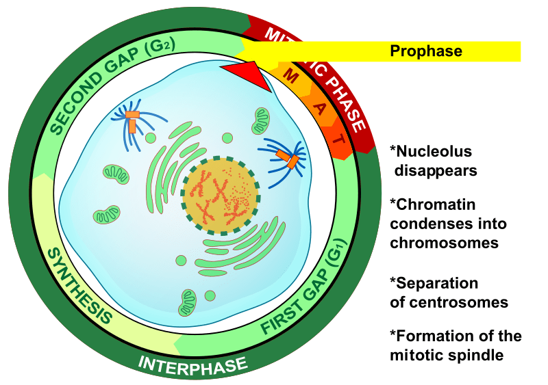
Prophase is the phase after interphase and the first step of the M-phase (cell division, i.e. mitosis and meiosis) of the cell cycle process and also the one where the chromatin condensation begins in order to manifest chromosomes formation. Both mitosis and meiosis consist of chronological phases that start at prophase (followed by metaphase, anaphase, and finally by telophase). In meiosis, prophase occurs twice (hence, the terms prophase I and prophase II).
- During mitotic prophase, the chromatin condenses, becoming more visible (chromatin condensation). The chromatin fibers condense to become distinct chromosomes that are visible when viewed under the light microscope at high magnification. Spindle formation begins and the nucleolus seems to disappear.
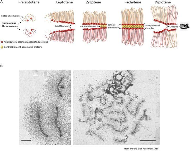
- In meiotic prophase, there are two prophase events: prophase I and prophase II. Prophase I takes place in the first meiotic division and is comprised of the following sub-stages: leptotene, zygotene, pachytene, diplotene, and diakinesis. Prophase II has no similar sub-stages and occurs in the second meiotic division. Nevertheless, both prophase I and prophase II are highlighted by the disappearance of the nucleoli and the nuclear envelope and chromatin condensation.
Etymology: Latin or Greek pró (before) + phase, phásis (“appearance”)
See also: cell cycle, cell division, prophase I, prophase II, prometaphase
Staining and Microscopy
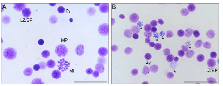
Chromosomes and cells are microscopic in nature. You can’t visualize a single cell, a DNA molecule or chromosomes with naked eyes. You need an aided vision for this exercise. When we talk about microscopy, the technique of staining also becomes integral. As the chromatins are condensed to chromosomes in the prophase stage and hereby the movement of chromosomes starts, it’s interesting to learn about this intricate yet interesting movement by visualizing it. We can stain both somatic cells and germinal cells (gametes) to visualize the prophase stage in mitotic division (prophase) and meiotic division (prophase I and prophase II) respectively. Some commonly used stains for visualizing chromosomal movements in various stages of cell divisions are:
-
Giemsa stain
The well-known G-banding pattern is based on Giemsa staining. The main constituents of Giemsa stain are methylene blue (MB), eosin, and Azure B. This stain can be used to stain nucleic acids as it a has specific affinity for the phosphate groups of DNA. This staining technique has been heavily exploited for making karyograms and the identification of many chromosomal aberrations. Giemsa stain isn’t just used for histopathological aspects but also for fungi, bacterial, and yeast studies. Nuclei are stained dark blue in color with Giemsa stain while the cytoplasm has a color ranging from pink to light blue.
-
Silver stain
This staining technique became more popular in recent times. This stain is also heavily used for visualizing the various stages of meiosis and mitosis including prophase.
Prophase in Mitosis
There is only one prophase in mitosis. In mitosis, prophase is the first stage of cell division.
Before the prophase of mitosis, the G2 phase of interphase occurred. And after the prophase of mitosis, metaphase occurs. We can credit an entire range of DNA binding proteins that aid the catalysis of the chromosome condensation process occurring in the prophase stage. Some of those DNA-binding proteins are cohesin and condensin.
- Role of cohesin: This protein ensures the formation of a ring-like structure that holds the sister chromatids together at the centromere.
- Role of condensin: This protein ensures the formation of multiple rings that coil and twirl around the chromosomes in very highly compact and condensed forms.
Another notable feature of prophase: The centrosomes move on the opposite sites to the poles. This leads to the establishment of the mitotic spindle as microtubule assembly occurs between the two centrosomes. This later accounts for the “pulling part” motion of the sister chromatids towards the opposite poles.
 |
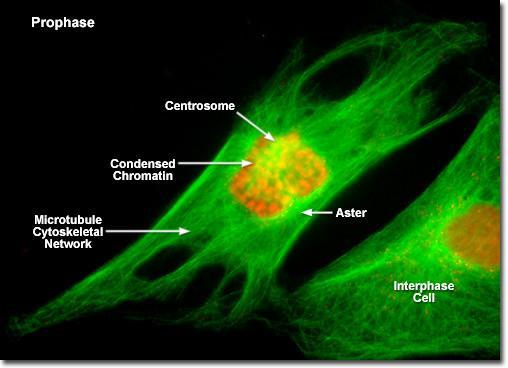 |
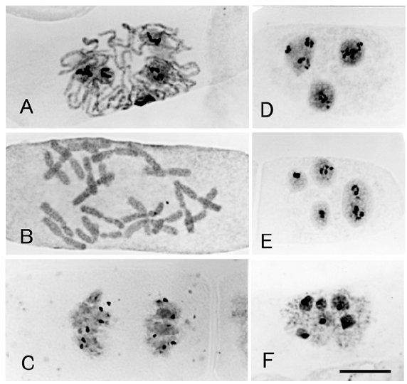
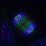
| Figure 4: These are some fluorescently labeled images for clearly understanding the various changes going on in the cell at the time of prophase. You should be able to understand the changes listed in the above sections after critically studying these images. Image Credits: Michael W. Davidson and The Florida State University. | |
Watch this vid about prophase in mitosis:
Prophase in Meiosis
There are two prophases in meiosis. In the germinal cells (gametes), after the completion of interphase, meiosis starts. The very first stage of meiosis is prophase I. There are some major differences between these two prophases. Let’s look at them individually.
Prophase I
This stage of meiosis occurs after the interphase. It is followed by two separate cell divisions (one in cytokinesis after telophase I and the other in cytokinesis after telophase II). Here, we visualize homologous chromosomes that separate in anaphase I.
There are multiple sub-stages in prophase I namely leptotene, zygotene, pachytene, diplotene, and diakinesis. There is the occurrence of crossing over event and chiasma formation in the prophase I stage. Crossing over of genetic material happens in the pachytene stage. The chiasma formation can be distinctly observed in the following stage, diplotene. Crossing over refers to the genetic recombination where two homologous chromosomes conduct the transfer of their genes between each other. These interesting steps ensure that new combinations of genes are formed when the two homologous chromosomes (one from the mother and the other from the father) merge for a short duration of time in prophase I. This also ensures that the offspring carries some new combination of alleles and genomic variation… ☺
Watch this vid about prophase I:
Prophase II
Prophase II occurs after telophase I. It is followed by only one cell division and there are no multiple/separate cell divisions unlike prophase I. Here, we visualize single chromosomes from which the sister chromatids separate in anaphase II. There are no sub-stages in prophase II. There is no crossing-over event or chiasma formation in prophase II.
Prophase I arrest
In human females, the preparation of the gamete cells begins as early as during the female fetal development. When a female young one is born, this preparation is arrested at the diplotene sub-stage of prophase I. This prophase I arrest ensures that the germinal cells (oocytes) don’t mature any further before the girl reaches puberty. Only when she reaches adolescence and hits puberty, then the levels of LH (luteinizing hormone) shoot up and meiosis resumes. At ovulation, an oocyte goes through the remaining sub-stages of prophase I, and then meiosis will stop again at metaphase II. Only during fertilization that the now secondary oocyte will complete meiosis. Otherwise, the cell will disintegrate at menstruation.

Differences in Plant and Animal Cell Mitotic Prophase
Apart from cytokinesis where plant cells form cell plate (cell plate later forms cell wall) and animal cells form cleavage to divide the parent cell into two daughter cells, a very important point to note when we talk about the difference in plant and animal mitosis is the mitotic spindle formation during prophase. Plant cells lack centrioles, which are present in animal cells. The centrioles organize into centrosomes, which in animal cells serve as the main microtubule-organizing center. Thus, the centrosomes dividing and migrating to opposite poles of the cell during prophase will only be observed in animal cells and not in plant cells. How do plant cells then organize their microtubules if they don’t have centrosomes? Plant cells have alternative microtubule-organizing mechanisms where the mitotic spindle can still form and organize similar to animal cells but without the centrioles. (Sanchez & Feldman, 2017)
Cell Checkpoints
There are many checkpoints in the cell cycle to regulate and overlook the proper cycle. Only when the cell assures that the previous steps were undertaken with precision and accuracy that the cell proceeds further. One of these checkpoints is G2-M checkpoint occurring in preprophase. This means that before the cell enters the mitotic phase, there is a definitive “G2-M Checkpoint” that needs to be crossed. The cyclin involved here is cyclin B.
Frequently Asked Questions on Prophase:
Chromosomes are duplicated during what portion of the cell cycle?
Answer: Interphase (Replicative phase/S-phase)
What happens during prophase?
Answer: Generally, the condensation of chromatin to chromosomes
What must happen before a cell can begin mitosis?
Answer: The genetic material must have been properly replicated as well as the cytoplasmic content must have increased enough for 2 daughter cells.
How many chromosomes will be found in each cell during prophase?
Answer: Chromosomes will double in number in prophase as this stage is preceded by S-phase (replicative phase in interphase).
During which phase of mitosis do the chromatids become chromosomes?
Answer: Anaphase in mitosis (Reason: Since the sister chromatids joined at centromere are pulled apart in the anaphase stage of mitosis, they technically can be called individual chromosomes here on at their respective poles!)
During which phase of meiosis do the chromatids become chromosomes?
Answer: The answer to this question isn’t simply anaphase as there are two anaphases in meiosis. The correct answer is anaphase II. (Reason: The homologous chromosomes separate in the anaphase I stage. It is only in the anaphase II stage that the sister chromatids separate and become individual chromosomes (the separation of homologous pairs of chromosomes is a noted feature of the anaphase I stage).
Crossing over occurs during which phase of meiosis?
Answer: Pachytene stage of prophase I of meiosis type of cell division (reductive division).
Answer the quiz below to check what you have learned so far about prophase.

References
- Ishishita, S., Inui, T., Matsuda, Y., Serikawa, T., & Kitada, K. (2013). Infertility associated with meiotic failure in the tremor rat (tm/tm) is caused by the deletion of spermatogenesis associated 22. Experimental animals, 62(3), 219–227. https://doi.org/10.1538/expanim.62.219
- Hizume, M. (2014). Chromosome analysis in Cupressus sempervirens L., Cupressaceae sensu stricto. Chromosome Botany, 9, 125-128.
- Grey, C., & de Massy, B. (2021). Chromosome Organization in Early Meiotic Prophase. Frontiers in cell and developmental biology, 9, 688878. https://doi.org/10.3389/fcell.2021.688878
- Greenstein D. Control of oocyte meiotic maturation and fertilization. 2005 Dec 28. In: WormBook: The Online Review of C. elegans Biology [Internet]. Pasadena (CA): WormBook; 2005-2018. Figure 1, Oocyte Meiotic Maturation and Egg Activation. Available from: https://www.ncbi.nlm.nih.gov/books/NBK19690/figure/A3722/
- Sanchez, A. D., & Feldman, J. L. (2017). Microtubule-organizing centers: from the centrosome to non-centrosomal sites. Current Opinion in Cell Biology, 44, 93–101. https://doi.org/10.1016/j.ceb.2016.09.003
©BiologyOnline.com. Content provided and moderated by Biology Online Editors.
