
Ribosome
n., plural: ribosomes
[ˈɹaɪbəˌsəʊm]
Definition: A minute, sphere-shaped particle composed of protein and ribonucleic acid (RNA) that serves as the site of protein synthesis
Table of Contents
A biological cell is composed of many components called organelles. These organelles serve their specific purposes to make the cell a wholesome living unit. An organelle is defined as a structure that is surrounded by lipid bilayers. By that definition, nucleus, endoplasmic reticulum, Golgi apparatus, mitochondria, and chloroplast (plastid) fall under the category of organelles whereas lysosomes, vacuoles, ribosomes, and nucleosomes may not be so because they lack such a lipid bilayer. Lysosomes and vacuoles are bound by a single membrane whereas ribosomes and nucleosomes do not have a membrane surrounding them.
There’s another way we can classify cell organelles. An organelle is a specialized subunit inside the cell that performs a specific function. In this case, there are two types of organelles:
(1) Membrane-bound organelles (includes double membrane-bound and single membrane-bound cytoplasmic structures). Examples: nucleus, endoplasmic reticulum, Golgi apparatus, mitochondria, plastids, lysosomes, and vacuoles.
(2) Non membranous organelles. Examples: ribosome organelle, nucleosome, spliceosome, vault, proteasome, DNA polymerase III holoenzyme, RNA polymerase II holoenzyme, photosystem I, ATP synthase, centriole, microtubule-organizing center, cytoskeleton, flagellum, nucleolus, stress granule, etc.
Now that we know the basic classification of cell organelles, let’s move ahead and learn about ribosomes in detail which is the main topic of this article. We will answer some common questions and doubts around the topic too, so keep reading…
Ribosomes Definition
The ribosome is a cytoplasmic structure that is minute and sphere-shaped. It is composed of protein and ribonucleic acid (RNA). As the famous ribosomes analogy to factories suggests, they serve as the site of protein synthesis; protein factories. Etymology: from ribonucleic acid and Greek: soma (body).
In 1955, ribosomes were first observed as dense particles or granules by George Palade with his electron microscope. He discovered these cell organelles which are the protein factories inside a cell. In 1958, the term “ribosome” was proposed by the scientist Richard B. Roberts.
 |  |
Basic Questions About Ribosomes
Let’s answer some basic questions first before moving ahead to the intricacies of the topic.
- What is a ribosome?
Answer: Ribosome is a cell organelle that’s cytoplasmic in nature and also called the protein factory of the cell. - What do ribosomes do?
Answer: Ribosomes serve as the synthesis factories for proteins. They translate the genetic message encoded in the form of mRNA to proteins. Proteins are made up of amino acids. So, when asked, “do ribosomes make proteins?”, the answer is yes! - Where are proteins made in a cell?
Answer: Since ribosomes are translating machinery, they carry out the essential process of protein synthesis inside a cell. The conversion of genetic code from DNA to mRNA to proteins isn’t feasible without the ribosomal function. For the central dogma of life to run, ribosomes play an indispensable role. - What are ribosomes made of?
Answer: Ribosomes are made up of 2 components namely ribosomal RNA (rRNA) and ribosomal proteins. - Where are ribosomes found inside a cell?
Answer: Ribosomes are either found freely floating in the cytosol or attached to the RER (Rough Endoplasmic Reticulum). - Do prokaryotes have ribosomes?
Answer: Yes, all prokaryotes have ribosomes in their cells. Without ribosomes, the cells won’t be capable of repairing damage, producing proteins needed for various essential pathways and processes, and maintaining life. - Do eukaryotic cells have ribosomes?
Answer: Yes, all eukaryotic cells have ribosomes like prokaryotic cells.

Now that we know how to define ribosomes and are clear about the basic knowledge, let’s move ahead and learn about some details.
Origin
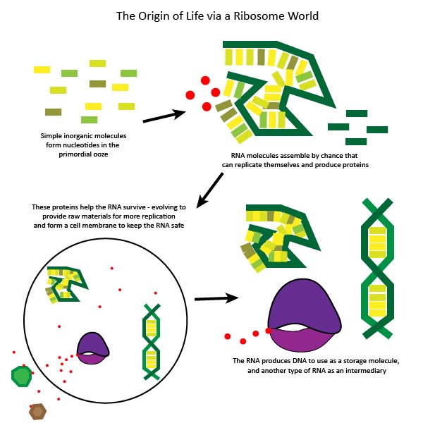
There are several opinions and hypothetical theories on the origin of ribosomes. There isn’t any one definite, full-fledged, evidence-backed hypothesis for the origin of ribosomes. Still, the best explanation provided to date resonates among many scientists. Some pointers to the theory are:
- It says that the ribosomes have evolved from some ancient “self-replicating complexes”.
- These complexes actually originated in the RNA world and have evolved to produce proteins.
- There are several studies that highlight that the early ribosomes were only rRNA complexes.
- Over the evolutionary time frames, they acquired the ability to synthesize proteins and peptide bonds. (Noller HF, 1992) (Noller HF, 2012)
- Ribosomes function in the new world as translational machinery.

Discovery
Here are the historical highlights of ribosomal discovery:
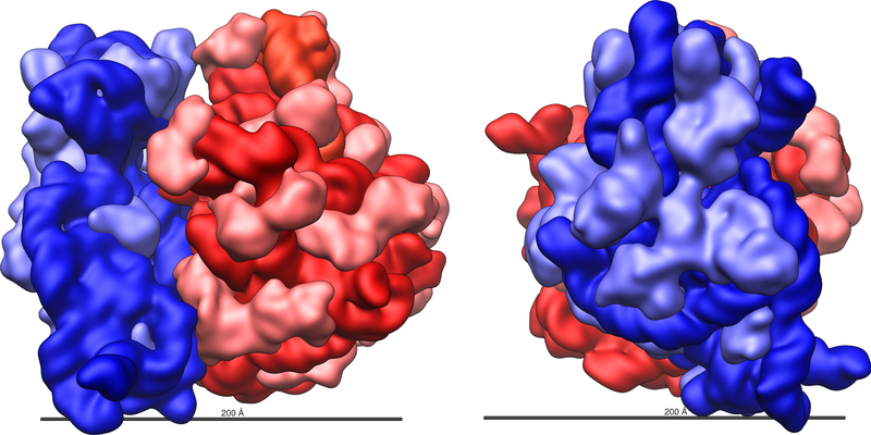
- In 1955, ribosomes were first observed as dense particles or granules by George Palade with his electron microscope. He discovered these cell organelles which are the protein factories inside a cell while studying the rat and chicken tissues. The nature of ribosomes as described by G. Palade was “granular, particulate and small-sized”. Another interesting fact noted down by him was the position of ribosomes. He said these particulate entities are present either floating freely in the ground matrix of cytoplasm or in close association with the endoplasmic reticulum membrane. He also estimated the diameter of these bodies as 100-150 Angstrom. Another point to note here is G. Palade discovered these entities but didn’t name them.
- For the discovery of ribosomes, Albert Claude, Christian de Duve, and George Emil Palade were decorated with the prestigious Nobel Prize in Physiology or Medicine in 1974.
- In 1958, the term “ribosome” was proposed by the scientist Richard B. Roberts.
- A detailed structure of ribosomes was given by Venkatraman Ramakrishnan, Thomas A. Steitz, and Ada E. Yonath. For this, they were later awarded the Nobel Prize in Chemistry in 2009.

Ribosome Structure
Ribosomes are made up of two basic components as discussed earlier: ribosome RNA (rRNA) and ribosomal proteins (R-proteins). There are various different types of proteins and their numbers differ from species to species. These 2 components are arranged in different constitutions in 2 different ribosomal units.
The two ribosomal subunits fit together and work as one to build proteins according to the genetic sequence held within the messenger RNA (mRNA). Ribosomes are typically composed of two subunits: the large and small subunits. They join as one during translation; together, they catalyze the translation of mRNA into a polypeptide chain during protein synthesis, and since their active sites are made of RNA, ribosomes are also referred to as “ribozymes.”

Bacterial ribosomes
- Size: Smaller than most of the ribosomes of eukaryotes (e.g. plants and animals); nearly 20nm in diameter. (Kurland CG, 1960)
- Composition: 65% rRNA + 35% ribosomal proteins
- Formed in the cytoplasm of bacterial cells.
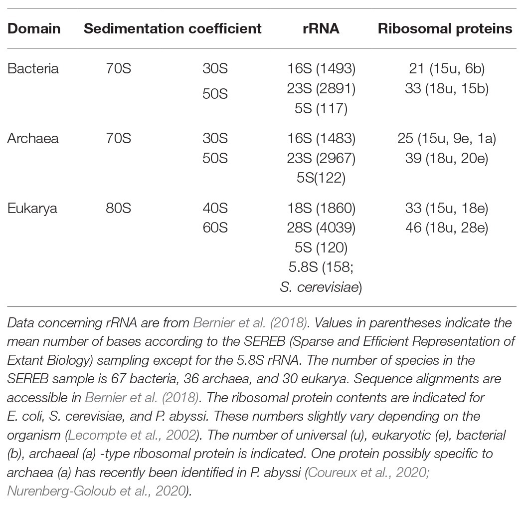
- The prokaryotic ribosome (70S) is made up of 50S (large subunit) and 30S (small subunit).
- In prokaryotes, the 30S ribosomal subunit contains the 16S rRNA whereas the 50S ribosomal subunit contains 5S rRNA and 23S rRNA.
- The numbers of nucleotides in each rRNA unit are:
- 16S rRNA subunit consists of 1540 nucleotides and is bound to 21 proteins.
- 5S RNA subunit consists of 120 nucleotides.
- 23S RNA subunit consists of 2900 nucleotides.
- 5S and 23S rRNAs are bound to 31 proteins.
Archaeal ribosomes
- The structure of archaeal ribosomes is structurally similar to bacterial ribosomes.
- They are also 70S and possess 2 subunits- 50S and 30S like the bacterial ribosomes. But they are similar to the eukaryotic ribosomes when compared at the sequence level. (Cullen, Katherine E., 2009)
Eukaryotic ribosomes
- Size: Bigger than prokaryotic ribosomes; nearly 25-30nm in diameter. (Wilson DN, 2012)
- The production of ribosomes involves both the cytoplasm and the nucleolus.
- The eukaryotic ribosome (80S) consists of 60S (large subunit) and 40S (small subunit).
- In eukaryotic cells, the 40S ribosomal subunit contains the 18S rRNA whereas the 60S ribosomal subunit contains rRNAs: 5S, 5.8S, and 28S.
- In eukaryotes, the semi-autonomous organelles like chloroplasts and mitochondria also have 70S ribosomes that resemble those of prokaryotes (e.g. bacteria). Because of this, it is suggested that these eukaryotic organelles have descended from their ancestral bacteria.
- The numbers of nucleotides in each rRNA unit are:
- 18S RNA consists of 1900 nucleotides and is bound to 33 proteins.
- 5S RNA consists of 120 nucleotides.
- 28S RNA consists of 4700 nucleotides.
- 5.8S RNA consists of 160 nucleotides.
- 5S RNA, 28S RNA, and 5.8S RNA are bound to 46 proteins.
Svedberg Unit
The subunits of the ribosomes are identified by their sedimentation rate represented by the Svedberg unit (S). The S units do not add up since they represent measures of sedimentation rate, not mass.
Below is a table that compares the structural components of prokaryotic and eukaryotic ribosomes.
Prokaryotic vs. Eukaryotic Ribosomes | |
|---|---|
| Prokaryotes | Eukaryotes |
70S (prokaryotic ribosome subunits)
| 80S
|

Plastoribosomes and mitoribosomes
- Plastoribosomes are ribosomes that are present in plastids (chloroplasts) of plant cells.
- Mitoribosomes are ribosomes that are present in the mitochondrial matrix. Mitochondria provide ribosomes with the precursors for the synthesis of proteins inside the mitochondria.
- Both are present only in eukaryotes as plastids and mitochondria are present only in eukaryotic cells.
- Type: 70S
- Similarity: These ribosomes share similarities with the prokaryotic ribosomes. Plastoribosomes are more similar to prokaryotic ones than mitoribosomes are to prokaryotic ones.

Making use of the differences
The structural and sequential differences in the prokaryotic and eukaryotic ribosomes are commercially exploited for the “development of antibiotics”. Scientists and pharmaceutical industries treat these differences as avenues for the creation of drugs that will specifically target 70S prokaryotic ribosomes of the pathogenic agent’s cells but won’t affect the 80S eukaryotic ribosomes of the patient’s cells.
CURIOSITY QUESTION: Do these antibiotics affect and target the mitoribosomes too? (As they are structurally similar to the prokaryotic ribosomes!!!)
ANSWER: No, as mitochondria are double membrane-bound organelles, hence antibiotics aren’t able to penetrate them and shut them off.

Common properties
- Core structure: is very similar in different types of ribosomes, although noticeably large differences exist in size.
- Ribosomal RNA organization: in tertiary structural motifs.
- The catalytic activity of the ribosomes: Carried out by the ribosomal RNA (rRNA).
- Ribosomal Structure Stabilization: by the ribosomal proteins (r-proteins) that reside on the surface.
High-resolution structure
The high-resolution structures of the ribosomes were published by studies from different scientific groups around the world. They were able to chalk out these structures to their atomic resolutions by studying them from some specific organisms. Here’s a list of the same.
Ribosome structures of some organisms | ||
|---|---|---|
| Subunit | Unit Description | Organism |
| 50S | Large prokaryotic subunit | Archaeon Haloarcula marismortui & Bacterium Deinococcus radiodurans |
| 30S | Small prokaryotic subunit | Bacterium Thermus thermophilus |
| 60S | Large eukaryotic subunit | Tetrahymena thermophila |
| 40S | Small eukaryotic subunit | Tetrahymena thermophila |
Functions of Ribosomes
Ribosomes perform vital functions inside cells. Ribosomes in plant cells and ribosomes in animal cells are organelles with huge responsibilities for the normal functioning of cells and life processes.
-
Translation
The ribosomes are described as the site of protein synthesis. Protein synthesis is the process of creating protein molecules. In biological systems, the major steps are amino acid synthesis, transcription, and translation. The ribosomes, though, are involved in the translation step. During translation, the amino acids are added by tRNAs and then are linked together in a specific order as specified in the mRNA transcript. So, when asked, “Where does translation occur?”… the answer is ribosome!
- Ribosomes are the protein factories. The translation process of converting mRNA code into protein happens because of ribosomes.
- Ribosomes hold the ability to decode the mRNA code.
- The ribosome moves along the mRNA template and reads each codon (made up of 3 nucleotides) of the mRNA.
- Then the ribosome pairs it with a specific amino acid. Amino acids are provided by an aminoacyl-tRNA.
- Aminoacyl-tRNAs are in possession of 2 things: first, a complementary anticodon on one end, and secondly, a specific, appropriate amino acid on the other end.
- Ribosomes have a unique ability to rapidly and accurately recognize appropriate tRNAs by putting their conformational proofreading ability at work.
Look at the picture below to understand the process of translation and the role of the ribosome in successfully completing it…

Figure 10: The process of eukaryotic translation is explained here. The internal ribosome entry site (IRES) is an important component of the translation initiation process. Peptidyl transferase aids the formation of peptide bonds and makes the protein chain elongation happen. There are 3 tRNA binding sites namely A, E and P on the ribosomes. Using the mRNA as a template, the ribosome traverses each codon, pairing it with the appropriate amino acid. This is done through interacting with transfer RNA (tRNA) containing a complementary anticodon on one end and the appropriate amino acid on the other. … or watch this video depicting translation
For proteins that need to be packed for transport (either within the cell or outside the cell), a signal peptide is the first to be synthesized. The signal peptide is produced by protein translation at the ribosome. This signal is an indication that the protein is for further processing in the endoplasmic reticulum (ER). When this signal is recognized by a signal recognition particle the ribosome translating the protein docks to the endoplasmic reticulum via the translocon. The ribosome, then, returns back to the translation of the protein. The chain continues to grow as the mRNA transcript is translated through the docked ribosome. The chain eventually makes its way into the ER through the translocon that spans across the ER membranes. The signal peptide is removed by a signal peptidase in the lumen of the ER. The nascent protein is folded in the ER by the chaperone proteins (e.g. ERp29, protein disulfide isomerase, BiP/Grp78, calnexin, etc.). The properly-folded protein is then packed into a transport vesicle to be shuttled to the Golgi apparatus where it would undergo maturation for transport along the cytoskeleton to other cytoplasmic organelles like lysosomes and peroxisomes or for secretion out of the cell.
-
Some basic questions:
- What does mRNA stand for? Answer: mRNA stands for messenger ribonucleic acid!
- What is mRNA made of? Answer: mRNA is made up of ribonucleotides!
- What does tRNA do? Answer: tRNA carries amino acids to ribosomes and helps in the proper incorporation of amino acids as per the codon in the mRNA.
- What is the ribosome in the human body? Answer: It’s a granular entity in all cells of the human body. If there were no ribosomes in the human body, no protein production would have taken place, and human life wouldn’t have been sustained.
-
Cotranslational folding
Another important role that ribosomes play inside living cells is the folding of proteins. This folding endows functionality to many proteins, hence making ribosomes furthermore important inside the cell.
-
Addition of translation-independent amino acids
Although we have always studied that the process of translation can’t happen without the presence of a coding mRNA strand. But there are cases where translation happens even in absence of mRNA strands. This is known as mRNA-independent protein elongation. A specific type of ribosomal quality protein aids in this process, (Rqc2). In this type of amino acid addition, the C-terminal of stalled proteins is extended. Alanines and threonines are the most common additions in such elongations.
Ribosome Locations
Ribosomes can be distributed in the cytosol or can be bound to the membrane of the endoplasmic reticulum. And thus, ribosomes are sometimes classified as either free or bound. The two types of ribosomes have similar functions and structures and are interchangeable. In fact, the bound ribosomes are attached to the ER transiently. They may come and go. They attach to the endoplasmic reticulum (via the translocon) when a signal peptide is synthesized by protein translation at the ribosome, and then recognized by a signal recognition particle.
-
Free ribosomes
In eukaryotes, the ribosomes may be classified as either ‘free’ or ‘bound’. Free ribosomes may be found suspended in the cytosol whereas bound ribosomes are attached to the endoplasmic reticulum (as such called rough endoplasmic reticulum). Free ribosomes’ function is for creating proteins, especially proteins that will function in the cytosol.
-
Membrane-bound ribosomes
Ribosomes found attached to the outer membrane of the endoplasmic reticulum are referred to as membrane-bound ribosomes. Bound ribosomes are involved in the synthesis of proteins that are to be exported or used within the cell membrane.
Biogenesis
Ribosome biogenesis refers to the biosynthesis of ribosomes. In eukaryotes, the sites of ribosome formation are the cytoplasm and the nucleus. The ribosome of eukaryotes is the 80S as opposed to the ribosome of prokaryotes, which is 70S. The 80S ribosome consists of a large subunit (60S) and a small subunit (40S). Each of these subunits is composed of ribosomal protein and rRNA(s). The ribosomal proteins are synthesized in the same manner as other proteins are produced, i.e. first by transcription within the nucleus and then moved into the cytoplasm for translation and maturation.
Mature ribosomal proteins are moved back into the nucleus, particularly in the nucleolus for ribosomal subunit assemblies, i.e. 60S or 40S assembly. As for the rRNA components of the 60S or 40S, they are produced in the nucleus.
In mammals, the rRNAs 18S, 28S, and 5.8S are transcribed in the nucleolus organizer region into a single unit pre-rRNA (referred to specifically as 45S pre-RNA) by the catalytic action of RNA polymerase I. The result is a large pre-rRNA made up of 18S, 28S, and 5.8S, which after processing would be released individually. As for the 5S rRNA, the genes encoding for it are transcribed into pre-5S rRNA by the RNA polymerase III. However, the pre-5S rRNA transcript is produced in the nucleoplasm, outside the nucleolus. Nevertheless, it finds its way to the nucleolus for the assembly.
To form the large subunit (i.e. 60S) of the ribosomal complex, 5S rRNA combines with 28S and 5.8S rRNA. 18S, in turn, forms the small subunit (i.e. 40S) by combining with the ribosomal proteins. These subunits would then be moved from the nucleolus into the cytoplasm for the assembly of a complete and functional 80S ribosome.
So, when asked about the site where ribosomes are made, they are made in the nucleus and at the cytoplasm.
Heterogeneous ribosomes
The heterogeneous nature of ribosomes helps in gene regulation. It has also been proposed to aid in the translational control of protein synthesis. It helps in the ribosomes’ structure maintenance and function.
Try to answer the quiz below to check what you have learned so far about ribosomes.

References
- George E. Palade – Facts. NobelPrize.org. Nobel Prize Outreach AB- https://www.nobelprize.org/prizes/medicine/1974/palade/facts/
- McQuillen, K., Roberts, R. B., & Britten, R. J. (1959). SYNTHESIS OF NASCENT PROTEIN BY RIBOSOMES IN ESCHERICHIA COLI. Proceedings of the National Academy of Sciences of the United States of America, 45(9), 1437–1447. https://doi.org/10.1073/pnas.45.9.1437
- Noller HF, Hoffarth V, Zimniak L (June 1992). “Unusual resistance of peptidyl transferase to protein extraction procedures”. Science. 256 (5062): 1416–9. Bibcode:1992Sci…256.1416N. doi:10.1126/science.1604315. PMID 1604315
- Noller H. F. (2012). Evolution of protein synthesis from an RNA world. Cold Spring Harbor perspectives in biology, 4(4), a003681. https://doi.org/10.1101/cshperspect.a003681
- Burma, D. P. (1992). RNA world and ribosomes. Current Science, 63(9/10), 550–560. http://www.jstor.org/stable/24094570
- Palade GE (January 1955). “A small particulate component of the cytoplasm”. The Journal of Biophysical and Biochemical Cytology. 1 (1): 59–68. doi:10.1083/jcb.1.1.59. PMC 2223592. PMID 14381428.
- Kurland CG (1960). “Molecular characterization of ribonucleic acid from Escherichia coli ribosomes”. Journal of Molecular Biology. 2 (2): 83–91. doi:10.1016/s0022-2836(60)80029-0
- Wilson DN, Doudna Cate JH (May 2012). “The structure and function of the eukaryotic ribosome”. Cold Spring Harbor Perspectives in Biology. 4 (5): a011536. doi:10.1101/cshperspect.a011536. PMC 3331703. PMID 22550233.
- Cullen, Katherine E. (2009). “Archaeal Ribosomes”. Encyclopedia of life science. New York: Facts On File. pp. 1–5. doi:10.1002/9780470015902.a0000293.pub3. ISBN 9780470015902.
- Schmitt, E., Coureux, P. D., Kazan, R., Bourgeois, G., Lazennec-Schurdevin, C., & Mechulam, Y. (2020). Recent Advances in Archaeal Translation Initiation. Frontiers in microbiology, 11, 584152. https://doi.org/10.3389/fmicb.2020.584152
- Alberts, Bruce; et al. (2002). The Molecular Biology of the Cell (4th ed.). Garland Science. p. 342. ISBN 978-0-8153-3218-3.
- Wilson, D.N. (2013). Ribosome-targeting antibiotics and mechanisms of bacterial resistance. Nature Reviews Microbiology, 12, 35-48.
©BiologyOnline.com. Content provided and moderated by Biology Online Editors.


