Control of Body Movement
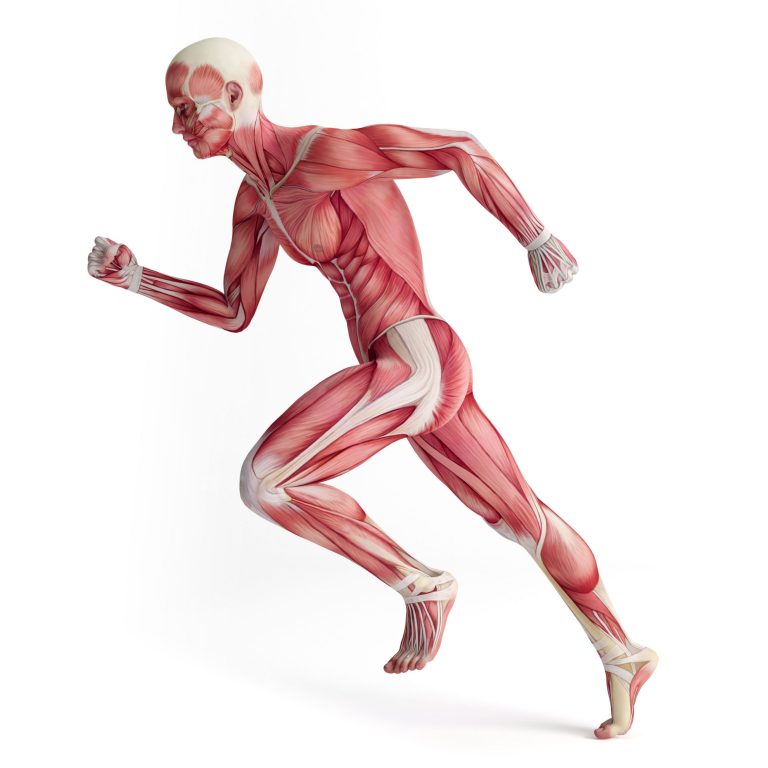
Neural activities are required to perform a movement.
Table of Contents
Motor Control Hierarchy
A motor program is the pattern of neural activities required to perform a movement is created and transmitted via neurons that are organized in a hierarchical manner. The program is continuously updated. Learning and skill can develop if the program is repeated frequently enough.
Voluntary and Involuntary Actions
Voluntary movement is accompanied by a conscious awareness of the action while the involuntary movement is not. All motor behaviors lie in a continuum and have both components in different degrees.
Local Control of Motor Neurons
Important in keeping the motor program updated by gathering information from local levels through afferent fibers.
Interneurons
Interneurons are synapses that integrate inputs from both higher centers and peripheral receptors.
Local Afferent Input
Afferent inputs to local interneurons bring information about the tension of muscles, movement of joints, etc. that in turn influence movements.
Length-Monitoring Systems
Changes in muscle length and rate of these changes are monitored by stretch receptors located within structures called muscle spindles that are present in modified muscle fibers called intrafusal fibers. (Rest of the fibers responsible for the force of a muscle are called extrafusal fibers.) Stretching a muscle fires these receptors while contraction of the muscle slows the firing of these receptors.
Afferent fibers from these receptors can take 4 pathways:
- Some fibers go back directly to motor neurons of the same muscle without the interposition of any interneurons and these arcs are called monosynaptic stretch reflex arcs.
- Some fibers end on interneurons that inhibit the antagonistic muscles and this is called reciprocal innervation.
- Some fibers activate motor neurons of synergistic muscles.
- Some fibers continue to the brainstem.
Alpha-Gamma Coactivation
The larger motor neurons that control the extrafusal fibers are called alpha motor neurons and the smaller- motor neurons that control the intrafusal fibers are called gamma motor neurons. These neurons are excited or coactivated at the same time to get continuous information about muscle length.
Tension-Monitoring Systems
Receptors located in the tendons called Golgi tendon organs monitor the tension on a muscle.
Withdrawal Reflex
If a stimulus activates flexor motor neurons and inhibits extensor motor neurons, moving the body away from the stimulus, it is called a withdrawal reflex. The effect is produced on the same side of the body where the stimulus arose (ipsilateral side) and an opposite effect may be produced on the other side (contralateral side) to compensate for any lost support due to the withdrawal. This is a crossed-extensor reflex.
Muscle Tone
Passive resistance of skeletal muscle to stretch due to some degree of alpha motor neuron activity and the viscoelastic properties of the muscle. Abnormally 1ight muscle tone called hypertonia can result in brief spasms, prolonged cramps or constant rigidity.
Maintenance of Upright Posture and Balance
Coordinated muscular activities support the skeleton and the upright posture. The afferent pathways of postural reflexes come from: (1) the eyes, (2) the vestibular apparatus (3) the somatic receptors. There are brain centers that coordinate this information and compare it with an internal representation of the body’s geometry. The efferent pathways are the alpha motor neurons to the skeletal muscles.
Walking
Walking requires the coordination of many muscles. Extensor muscles are activated on one side to support the body’s weight and the contralateral extensors are inhibited by reciprocal inhibition to allow the nonsupporting limb to flex and swing forward.

You will also like...
Animal Water Regulation
Animals adapt to their environment in aspects of anatomy, physiology, and behavior. This tutorial will help you understa..
Plant Water Regulation
Plants need to regulate water in order to stay upright and structurally stable. Find out the different evolutionary adap..
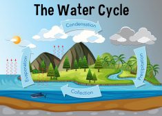
The Water Cycle
The water cycle (also referred to as the hydrological cycle) is a system of continuous transfer of water from the air, s..
Adaptation Tutorial
Adaptation, in biology and ecology, refers to the process or trait through which organisms or the populations in a habit..
Plant Biology
Plantlife can be studied at a variety of levels, from the molecular, genetic and biochemical level through organelles, c..
The Origins of Life
This tutorial digs into the past to investigate the origins of life. The section is split into geological periods in the..